Viewing entries tagged
breakthroughs

It’s in the Genes: Advances in Genome-Editing Tools

Comment

It’s in the Genes: Advances in Genome-Editing Tools

by Esha Ghai

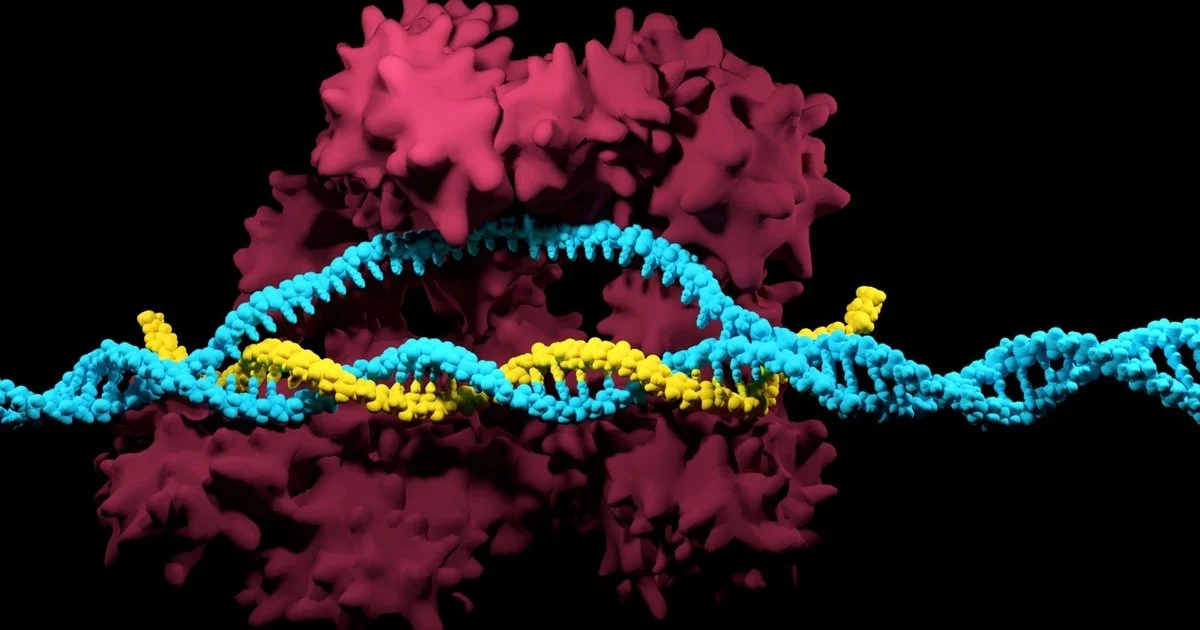
We have long been told that our DNA is unchangeable, often encouraging us to blame our flaws and accredit our talents to genetics. However, the introduction of genome-editing gives scientists the ability to alter genetic material and effectively change the basis of an organism. Genetic material can now be added, removed, or mutated at any specified location in an organism, allowing for cures to genetically inherited diseases and a wide range of illnesses. A recently discovered microbial ribonucleoprotein with such genome-editing capabilities is the CRISPR/Cas9 system. 

CRISPR/Cas9, short for “clustered regularly interspaced short palindromic repeats and CRISPR-associated protein 9”, was naturally developed by bacteria as a defense mechanism against invading viruses. As a part of this mechanism, bacteria confiscate fragments of DNA from invading viruses, and use the fragments to construct CRISPR arrays [1]. CRISPR arrays give the bacteria the ability to recognize identical or similar viruses, so if a similar virus attacks again, the bacteria produces RNA segments from the CRISPR array to specifically attack the virus’s DNA [1]. Cas9, an enzyme, is then used to cut apart the DNA, essentially killing the virus [1]. Scientists in the medical community are now adapting this gene-manipulation system to be used in cells in other organisms, including humans. The genome-editing tool has unlocked the potential to combat human genetic diseases previously deemed untreatable. A more cost-efficient, advanced, and precise method of gene-editing, CRISPR/Cas9 is revolutionizing the face of medicine as we know it [1].

Fascinated by the properties of CRISPR/Cas9 and microbes, Dr. Xue Gao, a professor of Chemical and Biomolecular Engineering at Rice University, currently investigates the applications of genome-editing and microbe-based natural product discovery. Her research “lies at the interface of chemical biology and biomolecular engineering with a primary focus on small- and macro-molecule discovery and their applications to human health, agriculture, and energy.” Dr. Gao’s contribution to the development of Statins, a pharmaceutical drug with lipid-lowering capabilities for those at risk of cardiovascular disease, piqued her interest in protein engineering. The frequent use of Statins in the medical society motivated Dr. Gao to continue synthesizing molecules to combat diseases. 

Her research also encompasses the use of enzyme-catalyzation (increase in the rate of a process due to an enzyme) for anticancer and antibiotic drug synthesis. Enzymes are powerful biological catalysts, accelerating chemical reactions by assisting in the formation of a transition state between the starting reagent and end product. Enzymes are also highly specific in which reactions it catalyzes, usually catalyzing only a single chemical reaction (or reactions very similar in nature), limiting side reactions and the ultimate formation of wasteful by-products [3]. According to Dr. Gao, whereas traditional chemical methods of synthesizing compounds require environmentally toxic solvents and harsh conditions, enzymes work well in moderate environments and are capable of producing a higher yield of 99%. As enzymes are proteins, extreme temperatures and pH levels result in denaturation, rendering the enzyme inactive. Therefore, enzyme catalyzation ability is enhanced at moderate conditions. In biological systems, a majority of processes utilize enzymes, from the transfer of CO2 in the body through the enzyme carbonic anhydrase, to breaking down sugars through the enzyme amylase. The development of enzymes to catalyze complex chemical reactions, called directed evolution, is a major focus of the Gao Laboratory. 

Dr. Gao ascribes “her love [for] exploring what has already been designed by nature” to the sheer intelligence of microbes and their methods of defense against viruses. “Microbes are super smart. They create protein encodings themselves to fight different types of environments.”  CRISPR/Cas9, another prominent example utilizing protein encodings and enzyme catalysis, has also attracted Dr. Gao’s interest. CRISPRs are specialized protein encodings, while Cas9 is an enzyme capable of cutting apart genetic material. Recently, Dr. Gao utilized the genome-editing capabilities of CRISPR/Cas9 to treat autosomal-dominant hearing loss. Autosomal-dominant diseases are inherited from an abnormal gene from one parent, even when the corresponding gene from the other parent is normal. Dr. Gao’s findings suggested that a targeted delivery of gene-disrupting agents such as CRISPR/Cas9 in vivo (within a living organism) can potentially treat such cases of hearing loss [2].

Dr. Gao’s research group is planning to do further work in CRISPR/Cas9 applications for a variety of human genetic diseases, and hopes to make breakthroughs in research through accommodating advanced computational tools and developing DNA sequencing technology (technology that determines the sequence of nucleotide bases). Dr. Gao envisions the next stage of her group’s research to be developing new enzyme-catalyzed reactions in the energy sector, particularly the production of biofuels, and combating food contamination in the agricultural industry. 

Dr. Gao’s current and future research aspirations in the field of human genetics and the energy sector serve as an inspiration for aspiring female scientists globally. She hopes that young females will follow their passion in STEM, and envisions a future where female scientists are not in the minority. Because of Dr. Gao and her vision, the Gao Laboratory is not only an embodiment of cutting-edge innovation in the future of enzyme-catalyzed reactions, but also the inspirational potential of female scientists. 

Works Cited

[1] What are genome editing and CRISPR-Cas9? - Genetics Home Reference - NIH. https://ghr.nlm.nih.gov/primer/genomicresearch/genomeediting (accessed Nov 8, 2019).

[2] Gao, X., Tao, Y., Lamas, V. et al. Nature. [Online] 2018, 553, 217-221. https://www.nature.com/articles/nature25164 (accessed Nov 8, 2019).
[3] Berg, J.; Tymoczco, John; etc. Biochemistry, 5th; W.H Freeman & Co: New York, 2002; 303-341.

Comment

Comment

Membrane Fusion And The Secret to Muscular Degeneration

by Lam Nguyen

In the human body, there are about 37 trillion cells. Each cell is made up of organelles that are busy creating proteins, digesting nutrients, and transporting different molecules or other organelles around. In order to facilitate the diverse functions different organelles assume, the plasma membrane, a barrier made up of proteins and lipids, helps compartmentalize different metabolic reactions to specific organelles and regions of the cell. Plasma membranes also play a critical role in the transportation of nutrients and signals between organelles through a process called fusion. A major topic within plasma membrane research, membrane fusion is a process in which two separate lipid membranes fuse into one single layer [1]. This process is essential in the creation of many organelles, such as the ER, a particular organelle that has fascinated Dr. James McNew, professor of biosciences, ever since he first came to Rice University.

Dr. McNew received his Bachelor’s of Science in Biochemistry from Texas A&M University, his Ph.D. in Pharmacology from UT Southwestern,  and he completed his postdoctoral fellowship at the Memorial Sloan-Kettering Cancer Center. His interest in cellular biology and membrane fusion started with his graduate work while researching how the peroxisome, an organelle responsible for various metabolic processes such as detoxification and cellular signaling, worked. This particular lab experience helped him realize that he had a passion for understanding basic molecular biology and “how different cellular components moved around the cell.” His burgeoning interest in basic cell biology followed him to his postdoctoral fellowship where his research centered around how proteins were moved, targeted, and carried to the Golgi apparatus, an organelle that processes and sends out newly synthesized molecules. Dr. McNew’s passion for cell biology and membrane fusion  has defined much of the work he has done here at Rice. His original research pertains to understanding membrane fusion in the endoplasmic reticulum (ER). The ER is a network of membranous tubules that is involved in protein and lipid synthesis1. Dr. McNew and Dr. Michael Stern, a fly geneticist and professor at Rice, observed how these tubules came together, but “the proteins involved were unclear.” They then hypothesized that the protein elastin, a protein present in connective tissue, played a significant role in ER membrane fusion. The two were able to confirm that elastin helps ER tubules come together, and they characterized the protein by studying it both in vivo (in a test tube) and back in fruit flies.

Dr. McNew also describes himself as a “basic cell biologist” and that his work is generally “far removed from something medicinal or therapeutic.” Work by clinicians on the elastin protein, however, has confirmed that elastin plays a role in physiological processes in humans. In fact, defects in the gene encoding for elastin play a role in hereditary splasic paraplegia (HSP), a group of genetic diseases where weakness and stiffness in the muscles gradually lead to muscular degeneration overtime.   This discovery has allowed Dr. McNew to connect his basic biology research to clinical applications. Alongside characterizing the mechanism of elastin in ER membrane fusion, Dr. McNew has also been deepening his understanding of how mutations in this protein can cause human diseases. 

Trying to understand how disruptions in the elastin and ER membrane fusion could cause the degeneration of muscles seen in HSP, Dr. McNew’s lab discovered that the loss of elastin function results in neurodegeneration which eventually leads to the deterioration of muscles. This explains how “people who have HSP lose motor control and their ability to walk.” Between adjacent neurons, small signaling molecules called neurotransmitters are transported  to send the electrical signal from one neuron to another. Dr. McNew observed that defects in elastin seen in HSP patients are somehow connected to the lack of neurotransmission. The lab was initially puzzled because they saw signs of muscle degeneration but not neurodegeneration. “Even though nerve damage is a known cause of muscle atrophy, conventional wisdom is that the nerve cells die first," Dr. McNew explains.  Consequently, Dr. McNew and his lab have begun investigating the cellular communication involved in this seeming paradox by learning about the other signalling molecules involved, specifically the torr kinase protein [2]. The McNew lab has been researching how the lack of neurotransmission is signalled by the torr kinase in order to provoke cell death in muscular tissue. 

Dr. McNew plans on building on the vast amount of data that his lab has obtained by further exploring other types of proteins that work with elastin and torr. Understanding these biochemical interactions will allow scientists to have a better picture of how elastin defects, lack of neurotransmission, and muscular degeneration are all connected to each other. The opportunity to discover new conclusions and to be on the forefront of scientific innovations is what often inspires undergraduates to get involved in research. When asked about if he has any advice for aspiring undergraduate researchers, he says that one has to be “okay with failure.” “It is very exciting and engaging to do something for the very first time. The feeling that you are the first person to do something is very addictive and keeps you going!” exclaims Dr. McNew. It is this excitement that will continue to inspire Dr. McNew in furthering his understanding of our cells and how they work.


Works Cited

(1) Daumke, O.; Praefcke, G. Structural Insights Into Membrane Fusion At The Endoplasmic Reticulum. Proceedings of the National Academy of Sciences 2011, 108 (6), 2175-2176.

(2) Loewith, R.; Hall, M. Target Of Rapamycin (TOR) In Nutrient Signaling And Growth Control. Genetics 2011, 189 (4), 1177-1201.


Comment

Comment

The Machines within Our Cells: Rewiring Genetic Circuits

by Gulnihal Tomur

Many people regard biology as simply trying to solve the mysteries of nature through observation.  A newly emerging field called synthetic biology bridges the gap between engineering and biosciences through reconstructing  already existing biological systems or fabricating entirely new ones by using artificial DNA synthesis technology. As synthetic biologists construct and insert manufactured genes in cells, they have two main goals in mind: either turn the cells into drug or biofuel factories or gain a better understanding of the roles different biological components play in naturally existing biological system. Synthetic biology allows scientists to reconstruct existing biological systems on a molecular level.

When asked to describe synthetic biology, Dr. Caleb Bashor of the BioScience Research Collaborative at Rice alludes to a famous quote from physicist Richard Feynman: “What I cannot create, I do not understand.” Dr. Bashor and his team attempt to rebuild intricate biological pathways to better comprehend their underlying principles. He refers to cells as micro-level machines — and “just like any machine, the cells have wiring inside them in the form of DNA and proteins.” Accordingly, Dr. Bashor’s approaches cellular systems from an engineering standpoint as he reconstructs these systems just like how an electrical engineer would rewire a machine to figure out how it works. “I am partly an engineer and partly a scientist, trying to understand how cells are organized so I can rewire them,” Dr. Bashor explained. 

Yet, Dr. Bashor did not start his scientific journey as a synthetic biologist; instead, he first occupied himself with biochemistry as an undergrad at Reed College. He was especially drawn to thinking about biology from the protein structure scale. At UCSF, he pursued the study of biological structures as a grad student from a perspective rooted in biophysics. Dr. Bashor got involved in synthetic biology at its early stages through a protein structure and function lab at UCSF, where they put different protein domains together to change the function of the protein as it was expressed in the cell. Dr. Bashor shares how they were essentially engaged in synthetic biology, yet did not know how broad the scope of their research was: “Eventually, people came along and told us ‘You guys are doing synthetic biology.’ And our reaction was ‘What’s that?’” As Dr. Bashor immersed himself more and more into this field, he dedicated his postdoc research at MIT to engineering transcriptional networks in eukaryotic cells. His current research at BioScience Research Collaborative encapsulates an even more ambitious approach as he and his team establish “synthetic regulatory circuits to reprogram the behavior of human cells.” Dr. Bashor and his team currently tackle mammalian regulatory circuits with potential applications that range from biosensing to cell-based therapy.

In order to fully understand Dr. Bashor’s work and its implications, we must first look into a natural gene circuit. Both eukaryotes and prokaryotes carry multiple genes in their genome, some of which get turned on and off depending on the external and internal signals cells receive. Since most proteins have complex structures, usually multiple genes are clustered together in a transcription unit. This then codes for the subunits of a single protein. The expression of these  genes are all dependent on a single promoter for their expression; the promoter serves as the site where RNA polymerase binds to DNA to initiate the transcription of the information embedded in DNA to mRNA. The so-called on-off switch of such transcription units is a specific DNA segment called an operator. Located either within or after the promoter, the operator is responsible for regulating when RNA polymerase can bind to DNA and start the transcription of the gene cluster. The promoter, the operator, and the clustered genes make up genetic circuits known as operons. 

When another protein called a repressor, which is the product of a regulatory gene separate from the operon’s gene cluster, is present in its functional state, it binds to the operator, thus physically blocking RNA polymerase from binding to the promoter. Internal and external factors, such as the presence of a specific molecule, influence the activity of the repressor by changing its 3D shape, which allows cells to switch on and off specific gene clusters based on environmental cues. Synthetic biologists take advantage of this knowledge as they introduce molecules in the cell which may turn on or off a repressor, add artificially designed circuitry into the existing systems, or isolate certain parts of a naturally occurring gene circuit through genetic modification. 

Mirroring and reconstructing such systems, however, still proves to be a real challenge despite the advancement in synthetic biology. One common problem scientists deal with is the instability of artificial gene circuits. These artificial circuits can break the natural balance of the metabolic systems of the cell. As the artificial gene circuits use the energy which would otherwise be used for growth and division, they hinder the cells from maximizing their fitness. Thus, any mutant cell in the cluster which did not accept the circuit outcompete the synthetically engineered cells. “The circuit becomes unstable evolutionary; the cells spit out the circuit over time in order to grow faster,” explains Dr. Bashor. Intrigued by this evolutionary process, he put his mind to engineering a circuit that doesn’t get spat out. He realized that he needed a device that would help him and his team monitor the evolutionary process of cells with artificial circuits in them. The result was eVOLVER — it enabled them to work with many circuits at a time. As an automated multi-culture platform, eVOLVER can control multiple parameters at once. It shows the cell growth and detects any disparities which may signal a mutant culture that spat out the circuit put into the system. “We can grow [the cells], watch the circuits break, and see where they recover their fitness as a result of the circuit breaking,” tells us Dr. Bashor. Ultimately, the device operates as a tool for being able to build circuits and then test their stability, which is a crucial factor if the circuit is desired to be stable for a long time so that, for instance, it can be used for the production of a chemical.

Another challenge scientists face presents itself in the complexity of multi-gene systems. Pleitropic genes, which influence two or more seemingly unrelated phenotypic traits, and gene clusters regulated by common factors hinder scientists who aim to understand the function of individual parts in such systems. Therefore, synthetic biologists turn to a technique called refactoring. “Refactoring is a term borrowed from computer science,” tells us Dr. Bashor, “somebody gives you a code that is really hard to work with, so you rewrite in a way that it is easier to change the parts individually and to see what you’re changing.” In a similar fashion, synthetic biologists “physically abstract genes away from their native genomic content.” (1) The new content in which scientists place the gene in question tends to be a simple plasmid. Not only does this new medium offer easier control over the effects and outcomes of the gene due to its simplicity, but it also allows scientists to quickly modify the new genomic content since putting a new piece of DNA in a plasmid is a relatively easy process. By eliminating the interference caused by the natural genomic context, synthetic biologists observe the role of specific genes in the whole system. Yet, Dr. Bashor warns us not to see interference as a sheer burden on the system. “You may make the regulation very simple, but then you lose something in the behavior. That’s a clue that the regulatory forces that are associated with that context are important.” In the end, he sees refactoring, and synthetic biology in general, as a trial-and-error process — synthetic biologists simplify and modify and rewire the systems they are curious about over and over again to master the role of individual parts, which then allow them to see the bigger picture more clearly.

There are many other obstacles in the synthetic biology scene scientists are still trying to overcome today. The inability to insert big chunks of DNA into cells, for example, is hindering scientists from taking full advantage of synthetic biology in cell therapy. Yet, Dr. Bashor sees the intersection of synthetic biology and cell therapy as one of the most promising developments to look out for in the future. As the trial-and-error process that is synthetic biology continues to ignite curiosity in current and future scientists, the field will offer more and more to the scientific community. From advancements in agriculture to biofuel production, the manipulation of biological systems has already enhanced our lives in ways we may not even realize, and it continues to help us finesse biological systems in ways we desire.

Sources

Synthetic Biology Explained. https://archive.bio.org/articles/synthetic-biology-explained (accessed Jan 20, 2020).

The Bashor Lab at Rice. http://bashorlab.rice.edu/index_large.html (accessed Jan 20, 2020).

Comment

Comment

The Solution Right In Front Of (or is it Inside?) Our Eyes

by Nithya Ramcharan

Antibiotics have for long been a prominent facet of medicine and are ubiquitous in households today due to their accessibility. At the earliest sign of a bacterial infection, we consume antibiotics with the confidence that it will be eradicated within a couple of weeks. However, this confidence has led to antibiotic abuse, a major issue in the medical field currently. For one, overusing antibiotics depletes the natural fauna populating certain organs like our intestines, making them more vulnerable to virulent bacterial strains. Naturally-occurring bacteria in our organs are a component of the immune system’s first line of defense and play a crucial role in immune system homeostasis and development [1]. Most importantly, antibiotic abuse has led to what some sources dub “the Antibiotic Resistance Crisis.” Antibiotics are often overprescribed when many of the infections’ pathogens are left unidentified, so out of caution, infections are assumed to be bacterial. 80% of antibiotics sold in the US are funneled into livestock for growth and prophylaxis [2].  As a result, bacteria are rapidly developing resistance to many commercial antibiotics and are returning more potent than ever. Bacterial genes can be transferred both to relatives and non-relatives through plasmids, circular DNA molecules in bacteria that can replicate independently of their host bacterial cell. This can confer resistance rapidly not only within a single population but also between different bacterial species [3]. Researchers are thus looking for alternatives to antibiotics to curb bacterial resistance and its catastrophic consequences.

One solution researchers have identified is a group of proteins occurring naturally within our bodies. Known as “antimicrobial peptides” (AMPs), these proteins are small, positively charged molecules that function within organisms as a natural immune response against microbes. Most commonly, AMPs can kill a bacterial cell through lysis; interactions between the positively charged clusters of an AMP and the negative charges in the phospholipid bilayer of a bacterium’s cell membrane form pores in the membrane, which ruptures the bacterium [4]. AMPs can also inhibit bacterial growth by targeting intracellular operations after permeating the membrane. They are present in organs in the human body most susceptible to infection such as the skin and the eyes [4]. Crucially, unlike antibiotics, bacteria have not developed solid resistance to AMPs, which is a key reason why AMPs offer a more attractive and effective method of treating bacterial infections. However, there is still little established research on how exactly AMPs neutralize bacteria. 

Professor Anatoly Kolomeisky of Rice University’s Department of Chemistry has been investigating the mechanisms of AMPs. His work started with the observation that antibiotic abuse can be attributed to people wanting to completely eliminate bacteria from their systems. His team developed a theoretical framework based on preexisting quantitative data to analyze how AMPs kill bacteria by considering bacterial growth as well as AMP entrance and inhibition rates into the bacteria as stochastic (randomly determined) processes. The model, in essence, consisted of a set of chemical reactions that would eventually lead to elimination of bacteria from the system. “We looked at the system as a sequence of states,” said Dr. Kolomeisky. “These states differed from each other only in the amount of AMPs.” In the occasion that the AMPs kill the bacterial cell, the corresponding state is removed. He called the model a “one-dimensional picture in a chemical space,” in the sense that it provided more of a black-or-white outlook: with certain factors, all the bacteria would either be killed or none of them would be killed. Although the model is simplified and every biophysical process is not accounted for, the model offers a convenient method for calculating the dynamics of the reaction, such as bacterial growth rate and bacterial clearance rate and the parameters that can modify these rates. Additionally, unlike other theoretical research on AMPs, this study takes into account the stochasticity of the processes of AMPs entering and inhibiting the cell [5]. This offers an effective means to further understand the mechanisms of AMPs that can be analyzed statistically and later compared with empirical data. 

Using the model, the team found out that the probability of inhibition and bacteria clearance rate have a positive relationship. Additionally, both processes of AMP entrance and inhibition of bacteria are equally important for AMPs to suppress bacterial growth. One finding is that higher heterogeneity in the AMPs facilitate faster entrance rates, while faster inhibition rates lead to lower heterogeneity among the types of AMPs. Controlling the heterogeneity in AMPs is key to balancing entrance and inhibition rates and thus achieving an optimal bacterial clearance rate [5]. Even though the stochastic model is simplified to fit the available information, the mean inhibition times predicted by the model are consistent with those observed in other studies [5]. 

Once these properties are understood, the applications of AMPs are numerous. They expand beyond inhibiting just bacteria and could suppress other pathogens. They can potentially be used in regulating cell division, facilitating wound healing, assisting in surgical healing, and suppressing cancer cells. AMPs activated by environmental factors such as pH have been developed for drug therapy. LL-37, an AMP found in humans, is one example of an AMP currently in commercial use. It has been identified to have bactericidal, anti-cancer, and anti-inflammatory properties [7]. Aside from medicine, they can be used in food production as natural food preservatives. One of them, pedocin PA-1 (produced by a diplococcus, a type of round-shaped bacterium), is used as a food preservative to prevent meat deterioration [6]. AMPs have also been identified as potential animal feed components to boost health and immunity in animal farming and aquaculture. AMPs with antifungal properties could be used as pesticides in the agriculture industry. 

However, much is left to discover about AMPs, as they are a diverse group of peptides that work differently depending on their environment; for example, some operate at the millimolar scale, while some require nanomolar concentrations to function. In addition, knowing how to optimize the entrance and killing rates is important in determining the ideal level of heterogeneity among the AMPs attacking the cell. Dr. Kolomeisky is advancing his current research on AMPs by investigating how using more than one type of AMP affects bacterial clearance. Implementing a “cocktail” mix, as he called it, of AMPs would kill bacteria more efficiently. Even though the rate of the reaction decreases as more types of AMPs are introduced, the probability of killing bacteria increases. He is also researching the dynamics of cancer development and cell cycle regulation, and could combine his research with AMPs to help better understand both processes. 

Once the mechanisms of AMPs are better known, they can revolutionize biomedicine. They can replace antibiotics in many medical treatments, thus mitigating the Antibiotic Resistance Crisis yet still allowing antibiotics to be used when needed. Reducing unnecessary usage of antibiotics will insulate them from the imminent threat of resistance developing in bacteria so rapidly. Beyond medicine, AMPs serve as better alternatives for inorganic chemicals used in food preservatives, growth promoters, and pesticides as they are naturally-occurring molecules that might be less likely to harm the organisms they aren’t programmed to target. In the nature of an archetypal journey of self-discovery, the solution to the antibiotic resistance crisis lay within us this whole time.


Comment

Comment

A Stressful Situation

by  Saksham Vashistha

Affecting nearly 80% of Americans, stress has become an integral part of our communities. However, while stress can affect our mental health, cells, too, experience physical stresses. However, while stress can affect our mood, physical stresses affect our bodies at the cellular level. Getting to the bottom of how cells respond to stresses is a useful tool in a wide variety of fields. Dr. Mike Gustin, a professor of biological sciences at Rice University, aims to understand stress responses in cells and their role in the treatment of cancer.

Dr. Michael Gustin’s passion for cellular biology developed during his time as a graduate student. While working in the Ching Kung lab at the University of Wisconsin-Madison, then-physiologist Dr. Gustin and his team were the first to discover that ion channels, special proteins that span the cellular membrane and help transport charged particles into and out of the cell, exist in yeast and bacteria. Now at Rice University, Dr. Gustin has made the transition to genetics, focusing on the impact of stress on cellular behaviors. His most recent works include collaborations with researchers across the Houston area, namely Dr. Erik Cressman, a chemist at MD Anderson. 

Dr. Gustin and Dr. Cressman worked together on an NIH funded project that investigates ways to treat liver cancer. Unfortunately, as Dr. Gustin points out, “for liver cancer, there is no good chemotherapy.” Oftentimes, cancerous tumors in the liver are inoperable and alternative treatments are required. However, Gustin and his team have found that “chemistry-driven exothermic reactions (heat-generating reactions) can ablate (remove) tissues in an increasingly controlled way.” There are a variety of different reactions that can be used to ablate cells. Dr. Gustin’s group decided to use a reaction between an acid and a base. By mixing the two solutions on the cell’s surface and allowing them to react, heat and a high solute concentration (osmolarity) are produced. This extreme heat combined with the high osmolarity is toxic to the cell and kills it.

As Dr. Gustin explains, “there is an ablation zone [on the cell’s surface] where the temperature is high enough to kill cells and there is a combination of not just heat, but also high osmolarity because of the salt that accumulates there.” Gustin’s group is interested in the peri-ablation zone, the area just around the region of high osmolarity.  It was hypothesized that the applied cellular stresses are followed by some types of cellular defense mechanisms or signaling pathways. These signaling pathways serve to protect the cell from physiological stresses, helping it survive the harsh ablation conditions. Understanding how these pathways work is key for ablation treatment in cancer patients. The goal is, as Dr. Gustin says, “to inhibit those pathways in a controlled way [to] control how much ablation actually happens.” Thus, by inhibiting certain cellular defense mechanisms, ablation in cancer cells can be modulated and used precisely during cancer treatment. 

One of the interesting finds during their investigation was that sometimes the stress applied to the cells prevented the cells from even responding. Upon further investigation, it was found that some of the genes that are activated for the stress response actually inhibited other stress responses. Dr. Gustin’s team attempted to understand these stress responses at the molecular level. By using computer modeling of known proteins that were involved in the stress response pathways, they were able to identify how the structures of the proteins were changed by the cellular stresses. Gustin’s team wanted to understand “what happens to the fluid around a protein and what happens when stresses combine around that protein.” Understanding what happens to the fluid around the protein can indicate what environmental changes the protein is experiencing. The alteration of the fluid condition can lead to altered protein structures. Changes in the molecular structure of proteins affects the cell’s ability to react properly to stress. This is because the structure of the protein is what determines its function. If the structure is altered in any way, the protein wouldn’t be able to carry out its function correctly. This molecular modeling approach can help scientists understand not only at the cellular but also the tissue and organ level of how stress responses operate in a mammalian body. Furthermore, ablation in tumor cells can be controlled via inhibition of the genes and subsequent proteins involved in the cellular stress response. This control over the cell’s genes can help make ablation more effective for liver cancer patients with inoperable tumors. 

Dr. Gustin’s investigation into harnessing the power of cellular stresses has the potential to change how physicians approach the treatment of cancer. By identifying mechanisms by which cancer cells resist ablation, Gustin’s work provides new insight into how physicians can directly alter cellular defense mechanisms to make them more vulnerable to treatment. While understanding the influences of stress on cells has made significant contributions in medicine, Gustin’s work can also have applications in other fields. Dr. Gustin has his eyes set on something greater. “I am really curious about … what happens during coral reef bleaching”, Dr. Gustin says with a glint in his eyes. Coral reef bleaching is a direct result of stresses (changing water temperatures) on the cells of the organism, causing them to lose the algae living in them. The algae are responsible for providing the majority of the coral’s energy, without them the coral starve and die.  Gustin hopes to use his knowledge of cellular stresses to “genetically engineer algae that stay in the coral” reagrdless of the water temperature in an effort to save an inumerable amount of coral around the world. Such an effort would help lead the effort to rebuild important coral reef ecosystems globally.

[1] Saad, L. Eight in 10 Americans Afflicted by Stress. https://news.gallup.com/poll/224336/eight-americans-afflicted-stress.aspx

[2] Fuentes, D.; Muñoz, N. M.; Guo, C.; Polak, U.; Minhaj, A. A.; Allen, W. J.; Gustin, M. C.; Cressman, E. N. K. A Molecular Dynamics Approach towards Evaluating Osmotic and Thermal Stress in the Extracellular Environment. International Journal of Hyperthermia2018, 35 (1), 559–567.

Comment

Comment

The Brains Behind Smart Wound Dressings

by Puneetha Goli


Did you know that nearly 1.35 million people die each year as a result of traffic crashes? Or that  injuries sustained from these accidents are the leading cause of death for people ages 5 to 29 (1)? Currently, those injured in accidents are required to wait for personnel to arrive and scope out the scene before any medical help from a wound care provider can be given. However, imagine if instead, in the time a person waits for emergency services, a “smart” band aid could be applied immediately to the wound, setting the wound until advanced medical follow-up is available. 

This is exactly what Dr. Swathi Balaji, an assistant professor of Surgery at Baylor College of Medicine, is working toward. Alongside Dr. Taiyun Chi, an assistant professor of electrical and computer engineering at Rice University, Dr. Balaji hopes to develop an artificial intelligence-assisted smart wound dressing to speed up the wound healing process. 

Dr. Balaji received a PhD in biomedical engineering from the University of Cincinnati after she fell in love with the applications of the field and recognized the impact she could make by combining her foundation in mechanical engineering with bioengineering.

“I came for a Masters in Mechanical Engineering at the University of Cincinnati,” said Dr. Balaji. “I loved the program, …[but] it became very obvious that we were [only] making small incremental changes.” Her Masters work served as a turning point in which she realized she wanted to enter a field where she could make a more long-term impact, leading her to pursue a PhD in bioengineering. “There was so much going on in terms of orthopedic research, biomaterials, regenerative tissue applications, so I was very fascinated with that,” said Balaji. 

The work that Dr. Balaji conducted as a PhD student on tissue engineering, as well as her subsequent work on understanding the regenerative mechanisms of fetuses has inspired her current projects at Baylor. The goals of her lab are to understand intracellular communications and how they translate into an individual’s response to injury, scarring, and wound healing. Through examining healing mechanisms in mouse models, Dr. Balaji has worked to establish a pipeline, ranging from basic investigations to more complex interactions with biomaterials to address her lab’s goals. 

Dr. Balaji’s interest in regenerative wound healing coupled with calls of action from the Defense Advanced Research Projects Agency (DARPA) to better understand what is occurring at the wound scene is what led to a collaboration between her and Dr. Chi and ultimately gave birth to the idea of this innovative smart wound dressing (2). 

Dr. Chi’s lab is working on the development of novel biology-microelectronics hybrid systems. The technology entails using advanced integrated circuits and microfabrication techniques - the same process of fabricating CPU, memory, and wireless transceivers chips in our cellphones and tablets (3) - to create miniature-sized electronic biosensors and bioactuators (4). Although the technology already exists, his sensors are unique in that they are one of the first that can record multiple biomarkers simultaneously and on a flexible membrane, ideal for human skin. Recognizing the potential interdisciplinary overlap, the two began discussing the possibility of integrating the research done in Dr.Balaji’s lab with Dr.Chi’s sensors in order to create a machine-learning system that would be capable of collecting information from a wound site and administering an individualized treatment. 

“It’s a very exciting project...,” said Dr.Balaji. “We started off with simple machine learning but the goal is to ultimately go into more advanced predictive modeling and make it smarter as we go on, and the dream would be ... to deploy this in the battlefield or when there is an emergency or when there is an accident - to be able to slap it on and let the wound healing happen right from the get-go.”

Currently, the project is in its initial stages. The teams have worked to perfect their respective independent technologies—Dr.Balaji’s team has developed their understanding of the science using animal models, while Dr.Chi’s team has refined the sensors and actuators. Now, they are currently working to integrate both of these components and apply them to a wound site. 

However, despite the preliminary nature of her work, Dr.Balaji already sees multiple avenues for the future of her research. One of her goals is to promote this niche field of science in an academic environment:  “I think our goal is to develop this new field of biology - machine learning microelectronics hybrid systems - either as a new course for undergraduate or graduate education or a new pipeline for promoting STEM-based education,” said Balaji. In addition to the educational angle, Dr. Balaji expressed an interest in getting the SMART wound dressing patented. In the future, she hopes for a world in which her SMART wound dressing could have a wide range of applications - from healing a scratch from a playground fall to something more complicated such as a burn wound. 

The potential of Dr.Balaji’s and Dr.Chi’s project has been recognized by the John S. Dunn Foundation Collaborative Research Award Program and the duo was awarded the 2020 John S. Dunn Collaborative Research Awards which was established to “fund projects with high potential for impacting human health”(5). “We were very excited..[and] we see a lot of potential,” Dr.Balaji said of winning the award. She believes the award will give her and Dr. Chi’s team the necessary resources to dedicate the time and effort to produce a final product that can be tested and examined. “I think at the current stage, we have all the different pieces of the puzzle, and over the course of the next two years, ...the Dunn Award is really going to help us bring our technology together. To come up with an idea that is received very well makes it a very, very rewarding experience.” 

References 

  1. Road traffic injuries https://www.who.int/news-room/fact-sheets/detail/road-traffic-injuries (accessed Feb 24, 2021). 

  2. Intelligent Healing for Complex Wounds https://www.darpa.mil/news-events/2019-02-06a (accessed Mar 3, 2021).

  3. https://www.lifewire.com/what-is-cmos-2625826

  4. https://www.nature.com/subjects/biosensors

  5. Funding Opportunities – Gulf Coast Consortia https://www.gulfcoastconsortia.org/home/research/funding-opportunities/ (accessed Feb 24, 2021).

Comment

Comment

X Marks the Spot: High-Specificity Alternatives to Small Molecule Drugs

by Jessica Cao

“Side effects may include headache, dizziness, nausea, and vomiting, and serious, sometimes fatal allergic reactions may occur.” Most common and modern-day pharmaceuticals, such as those seen in medical advertisements, are classified as small molecule drugs (SMDs). These are defined as drugs with low molecular weight and are “capable of modulating biochemical processes to diagnose, treat, or prevent diseases”. [1] The nature of SMDs makes them particularly attractive in the pharmaceutical industry, as they can be easily manufactured and regulated compared to more complex compounds. This ease and convenience in turn lowers costs, allowing them to be more accessible and affordable for patients. [2] Unfortunately, the simple composition of SMDs typically results in a litany of unwanted side effects due to their inability to target specific protein receptors.The physiological dangers of these side effects have spurred the scientific community to discover other, potentially safer methods of drug delivery. 

Among these researchers is Dr. Jerzy Szablowski, an Assistant Professor of Bioengineering at Rice University. Dr. Szablowski’s research focuses on neurotherapeutics, particularly the development of technologies that can noninvasively monitor and interact with the brain.3 One of the projects at the center of his research is gene therapy. Dr. Szablowski believes that this is a distinctly promising method of drug delivery “because it can be separated into different layers of specificity.” He states that, unlike small molecule drugs, where “everything is encoded within several atoms of the molecule”, gene therapy can be optimized for specificity and modularity. Gene therapy involves the delivery of a new gene to a cell through a viral vector and a promoter, which is a DNA sequence that helps facilitate the production of the protein encoded by the gene. This type of multifaceted system is advantageous because each component can be modified for different uses. Since the purpose of the viral vector is to deliver the gene (the therapeutic component), the vector can be modified to treat specific parts of the body without altering the treatment. Furthermore, Dr. Szablowski elaborates that “the promoter [of the gene] determines subtype, [which can] provide specificity.” Ultimately, the ability to modify each  component of gene therapy without affecting the others yields greater specificity for each drug. Greater specificity allows for the avoidance of side effects resulting from non-specific binding between the drug and body receptors that are unrelated to the disease being addressed. However, assessing neurological illnesses to determine necessary treatment is often difficult, as brain biopsies are highly invasive and risky. 

In order to gain a better understanding of the pathology of neurological disorders and the effects that they have on specific parts of the brain, Dr. Szablowski and his lab have employed a method of controlling and monitoring brain tissue via ultrasound. This noninvasive technique involves focusing ultrasounds on a specific part of the skull to open the blood-brain barrier (BBB) by a few millimeters. As Dr. Szablowski describes, the ultrasounds are delivered in conjunction with “a special contrast agent that is FDA-approved, [which is] basically a bubble that is encased in a lipid. Because this contrast agent has gas, it can actually become compressed or [expanded].” He further specifies that ultrasounds are essentially “an expansion and compression of a mechanical wave over time”, which pushes apart blood vessels in the brain, causing the [impermeable] junctions of the BBB to separate, and the endothelium (a thin membrane) in the brain to become more permeable. During this stage, a patient’s blood can be drawn, and a simple blood test can be used to assess their neurological condition.4 This procedure can alternatively be used to deliver therapeutic nanoparticles and proteins such as antibodies to specific parts of the brain. Although the concept of opening the BBB may sound risky, Dr. Szablowski assures that “several hours post-application, the impermeable junctions basically close up, and they become normal again.” Furthermore, little to no side effects have been observed apart from minor brain inflammation, which is easily treated with the steroid dexamethasone. Dr. Szablowski believes that this method of focused ultrasound can be used independently or in conjunction with gene therapy to ultimately become a common method of highly specific drug delivery to the brain. 

Dr. Szablowski also hopes to expand on his current projects to discover novel methods of low-cost drug delivery, such as non-genetic therapy. He mentions that non-genetic therapy involves the “delivery of proteins that actually produce the drug on site, and the production of the drug on site allows it to be specific.” In other words, the biological site(s) involved with a disease are the only sites being treated, with other parts of the body remaining unaffected. Furthermore, the Szablowski lab has recently begun the Therapeutic Improvement Project, which will potentially simplify the arduous and expensive process of drug development. Dr. Szablowki says that currently, when developing a drug, “you first have to make a cell model of the drug, [then] find what receptors are responsible for the disease, then test [using a] tissue culture and mouse or larger animal, and if you're lucky and pass through all those stages, you can [use on] humans.” Unfortunately, because animals and humans are biologically different, a drug can affect each organism differently, and projects often remain “stuck” at the animal model phase. Dr. Szablowski and his lab are hoping to bypass the many obstacles of drug development by creating “a single drug [that] can treat multiple diseases by just delivering to single cells.” 

Ultimately, Dr. Szablowski hopes that his research will be applied to a number of diseases—most notably neurological ones such as Parkinson’s, epilepsy, and PTSD—as opposed to a specific illness. Many of these diseases are currently difficult to assess, monitor, and treat in real time, but Dr. Szablowski believes that these minimally invasive, yet highly specific methods will eventually allow the development of therapeutic treatment with little to no side effects. 


[1] Ngo, H. X.; Garneau-Tsodikova, S. What are the drugs of the future? https://www.ncbi.nlm.nih.gov/pmc/articles/PMC6072476/ (accessed Feb 24, 2021). 

[2] Gurevich, E. V.; Gurevich, V. V. Therapeutic potential of small molecules and engineered proteins. https://www.ncbi.nlm.nih.gov/pmc/articles/PMC4513659/ (accessed Feb 24, 2021). 

[3] Szablowski, J. Jerzy Szablowski: The People of Rice: Rice University. https://profiles.rice.edu/faculty/jerzy-szablowski (accessed Feb 24, 2021). 

[4] Miller, B. Targeting ultrasound for noninvasive diagnosis of brain cancer: The Source: Washington University in St. Louis. https://source.wustl.edu/2020/08/targeting-ultrasound-for-noninvasive-diagnosis-of-brain-cancer/ (accessed Feb 24, 2021).


Comment

Comment

The Mighty Might-ochondria

by Pavithr Goli

Famously known as the “Powerhouse of the Cell”, the mitochondria is vital as it provides the human body with the necessary energy to function on a day-to-day basis. In addition to this main role, researchers have recently discovered other mitochondrial functions critical to human survival such as interacting with pathogens during infections and metabolizing cholesterol (1). These often overlooked features of mitochondrial function have intrigued Dr. Natasha Kirienko, an Associate Professor in the Department of Biosciences at Rice, who has been studying mitochondrial surveillance pathways and damaged mitochondria. Furthermore, Dr. Kirienko has spent time studying how both can be utilized in anticancer therapeutics, which can potentially change the approach taken to treat cancer, saving numerous lives in the process.

Dr. Kirienko joined Rice University in 2015 after she earned a $2 million grant from CPRIT (Cancer Prevention & Research Institute of Texas). After earning her undergraduate and graduate degrees in biochemistry and biology in Russia, Dr. Kirienko came to the United States to work on her Ph.D. After receiving her Ph.D. in molecular biology from the University of Wyoming, Dr. Kirienko worked as a postdoctoral research fellow at the Massachusetts General Hospital and Harvard Medical School before joining Rice to continue her groundbreaking research on mitochondria. Dr. Kirienko developed her fascination for mitochondria during her time in graduate school where she was interested in why “mitochondria had lower efficiency in leukemia” and why “they had a poor ability to convert electrons into ATP.” 

As someone who has “always [been] interested in identifying fundamental principles of why things are working,” Dr. Kirienko was curious about the effect of disease on mitochondria and, “the biochemical basis of the cancer cells /cell lines.” This interest in studying the pathogenesis of mitochondria propelled her to begin conducting research and investigating the response of damaged mitochondria and the strategies these damaged structures employ to recover when pathogens impact their function. 

Dr. Kirienko spent years dedicated to developing a greater understanding of mitochondria with the hope that a new therapeutic method to fight cancer could be developed. This interest in understanding fundamental principles led Dr. Kirienko to focus her research on one of the most fundamental and basic organisms that exist: Caenorhabditis elegans or C. elegans (2). A roundworm commonly used in scientific research as a model organism, C. elegans has been instrumental in understanding the various molecular and genetic mechanisms the human body employs when responding to diseases like cancer (2). 

Using this model organism and previous literature as a guide, Dr. Kirienko has been able to observe novel pathways in mitochondria and find drugs that induce mitochondrial damage, pushing “cancer cells [to] undergo cell death like paraptosis or apoptosis.” The goal of Dr. Kirienko’s current approach is to apply this novel method of inducing cell death on ''different cancer cell types” or combine it with current anticancer drugs in a synergistic manner to create highly selective treatments capable of killing cancer cells and sparing healthy ones. This approach will help reduce the side effects commonly seen in cancer treatment while maintaining the treatment’s effectiveness.

To further her research, Dr. Kirienko uses an interdisciplinary approach by working with physical chemists and doctors to help ensure that her novel cancer therapy will be applicable beyond C. elegans. She hopes that physical chemists can optimize the drugs that Dr. Kirienko found by “synthesizing and improving the physical structure of the drug.” After further testing on mice and cell cultures, Dr. Kirienko intends to expand her research and consult the help of physicians in a potential clinical study.

Along with her cancer research, Dr. Kirienko has expanded her lab’s focus on other diseases caused by poor mitochondrial health such as Alzheimer’s disease. Specifically, she and her lab are trying to understand whether improving mitochondrial health may be beneficial for neuronal function. In order to understand this mechanism further Dr. Kirienko is attempting to use various biomolecules to avoid cellular death by activating mitochondria and “[finding] a way to help mitochondria fix themselves quicker.” 

Furthermore, Dr. Kirienko and her lab are attempting to observe mitochondrial reactions to infections. Using C. elegans as their model organisms, they are interested in “looking at basic toxins that damage mitochondria.” One specific group of toxins that they are investigating are siderophores, which are “small molecules with a peptide side chain… [that] are secreted in the body and release iron.” During a bacterial infection, the bacterial pathogen uses its siderophore to absorb iron from the host, enabling the bacteria to grow and infect the cells. The mitochondria are the site of electron transport chains (ETCs), which contain several iron-rich proteins that help generate energy for the cell; unfortunately, when “iron is stolen, the mitochondria get damaged.” (3).To better study this mysterious process, Dr. Kirienko and her lab have “developed a cell culture model where [they] can assess the pathogenicity of bacteria.” They aim to use this newly developed model to elucidate the reactions and mechanisms that allow pathogenic bacteria to hijack cellular mechanisms.

Beyond discovering mitochondrial pathways and reactions to infections, Dr. Kirienko's lab is eager to explore "mitophagy or mitochondrial turnover, applied to cancer… [and] study more mitochondrial pathways.” She hypothesizes that this research can provide key information in finding ways to strengthen mitochondrial health so that mitochondrial cells can effectively withstand deadly pathogens and diseases, ultimately leading to longevity in human health. 

Dr. Kirienko and her lab have been working on some groundbreaking research for the past five years. Using novel methods and ingenuity, Dr. Kirienko has already made major strides in the field of cancer therapy and molecular biology. Through her current projects and the future endeavors that she hopes her lab can make, Dr. Kirienko intends to understand more about the powerhouse of the cell and use it to develop influential cancer therapies. 

Works Cited:

(1)Wallace, Douglas C. “Mitochondria and cancer.” Nature reviews. Cancer vol. 12,10 (2012): 685-98. doi:10.1038/nrc3365  

(2)Why Study C. Elegans?, Dr. Lundquist, 01/19/21, www.people.ku.edu/~erikl/Lundquist_Lab/Why_study_C._elegans.html

(3) Montine TJ, Morrow JD. Fatty acid oxidation in the pathogenesis of Alzheimer's disease. Am J Pathol. 2005 May;166(5):1283-9. doi: 10.1016/S0002-9440(10)62347-4. PMID: 15855630; PMCID: PMC1606384.


Comment

Comment

A Memorable Discovery: Tracking Alzheimer’s with Light

by Jessica Cao

Every 65 seconds, someone in the United States develops Alzheimer’s disease [1]. Alzheimer’s disease is the most common form of dementia, a general disease marked by a decline in cognitive function severe enough to interfere with daily function. Alzheimer’s in particular is characterized by cognitive impairment and memory loss, and is most prevalent in people ages 65 and older [2]. These symptoms associated with the disease appear to be similar to aging, but are not part of the natural aging process [2]. There is currently no cure for Alzheimer’s, with current drugs only treating the symptoms (memory decline, confusion and disorientation), but significant developments have been made in the past few years towards understanding the disease. Among those in the research community advancing Alzheimer’s research is Dr. Angel Martí, an Associate Professor of Chemistry, Bioengineering, and Materials Science and NanoEngineering at Rice University. Dr. Martí’s lab is focused on the treatment and diagnosis of amyloid-related diseases such as Alzheimer’s and Parkinson’s disease, both of which are centered around the Beta Amyloid Hypothesis. 

The exact cause of Alzheimer’s is unknown, but several hypotheses have been formed in the past decades that may explain how the disease develops, most notably the Beta Amyloid Hypothesis. Beta amyloid (Aβ) is a sticky, hydrophobic protein found naturally in the brain in the form of monomers, its smallest molecular form. The monomers are derived from a larger protein known as Amyloid Precursor Protein (APP), whose original function is unknown. In patients with Alzheimer’s, Aβ monomers begin to aggregate and form Aβ oligomers; these oligomers may then aggregate further to form Aβ plaques, the most pathogenic form of Aβ [3]. Although Aβ plaques have been the main focus of Alzheimer’s research, Dr. Martí believes that understanding the development of Aβ oligomers—the aggregated form of Aβ proteins—is equally critical. Unlike plaques, Aβ oligomers are soluble in the blood and tissue fluid, allowing them to diffuse to different parts of the brain. This is particularly damaging since the oligomeric form of Aβ kills neurons as it diffuses. As Dr. Marti describes, “if Aβ is toxic [and] kills neurons, then as neurons start to die in patients with Alzheimer’s, the symptoms associated with [Alzheimer’s disease] will start.”

Taking this into consideration, Dr. Martí and his lab have created a ruthenium-based fluorescent complex, based on a concept known as fluorescence anisotropy, which tracks the development of Aβ oligomers in the brain. Fluorescence anisotropy is centered around the polarization of light, where “molecules can only absorb light that is in the direction of their transition dipole moments”. In other words, molecules can only absorb polarized light if they are oriented in the right direction, and will then rotate and emit light, resulting in a different polarization. However, since the molecules are extremely small, their high rotation speed results in the emission of light in multiple directions, depolarizing the light; the molecules can only emit polarized light in a specific direction when rotated more slowly. “Using fluorescence anisotropy, what you can measure is something that is proportional to how fast the molecules can rotate in solution.” The speed of the molecules is measured using a probe, which can bind to molecules if they are large enough. Aβ monomers are very small and are unable to interact with the ruthenium complex, “but once Aβ monomers start coalescing into bigger oligomers, then the probe can bind to the oligomers. And then, that probe that was rotating very, very fast before now rotates at the same rate as the big oligomer.” Using this complex, Dr. Martí and his lab may eventually be able to track the development of Aβ monomers into oligomers in Alzheimer’s patients by monitoring the changes in the speed that the probe rotates.

What can this be used for? The ruthenium-based fluorescent complex is still being refined and developed, but Dr. Martí says that it will eventually be used to “visualize or track [Aβ aggregation] in real time,” which will allow his lab to “test different drugs or molecules that might interact with these oligomers, and either break them apart or inhibit their formation”. Beyond its various applications, Dr. Martí believes that fluorescent complex may be further improved or by modifying the metal complex for increased binding efficiency, and possibly even modifying it to bind to proteins of other diseases. While this complex is not a cure for Alzheimer’s, it is undoubtedly a significant development in tracking the impact of potential cures. 

Works Cited

[1] Alzheimer’s Association. Alzheimer’s Disease Facts and Figures. https://www.alz.org/alzheimers-dementia/facts-figures (accessed Nov. 15, 2019).

[2] NIH National Institute on Aging. What is Alzheimer’s Disease? https://www.nia.nih.gov/health/what-alzheimers-disease (accessed Nov. 15, 2019). 

[3] Alzheimer’s Association. Beta-amyloid and the Amyloid Hypothesis. https://www.alz.org/national/documents/topicsheet_betaamyloid.pdf (accessed Nov. 15, 2019).

[4] Rice University. Angel Martí Group at Rice University. https://martigroup.rice.edu/(accessed Nov. 15, 2019).

Comment

Comment

One Mouse at a Time: Answering the Alzheimer’s Conundrum

by Mabel Tang

Alzheimer’s disease is the sixth leading cause of death in the United States [1]. One third of American seniors die with Alzheimer’s or dementia, and 5.8 million Americans live with the disease [2]. This number is projected to reach 14 million by the year 2050 [3]. Despite the large numbers of people affected by the disease, it has no cure. 

What we do know, however, is that there are several causes of Alzheimer’s disease. Studies suggest that the accumulation of proteins such as alpha-beta peptides and the tau protein in the brain contribute to the development of Alzheimer’s [4]. These aggregated proteins most likely disrupt neural activity and other circuits that control and support learning and memory, resulting in the death of neurons. Alzheimer’s can also be hereditary, although deterministic genes, or genes that directly cause disease, make up a very small percentage of cases [5]. On the other hand, Alzheimer’s risk is increased through head injury, diabetes and obesity, and age [6]. 

Dr. Joanna Jankowsky, a professor of neuroscience, neurology, neurosurgery, and molecular and cellular biology at Baylor College of Medicine, is seeking to better understand this devastating disease by modeling it with specially engineered mice. When Dr. Jankowsky first started her Ph.D at the California Institute of Technology, she was studying models of learning and memory. However, after working with a neurosurgeon on models of epilepsy, she found what she considers “the ground truth of the human disorder... You have a whole population of patients who want research on their disease because it’s so traumatizing for their life and their families’ lives. [My research] became much more grounded in something that seemed worthwhile. I really liked learning and memory, but I loved doing things that were clinically relevant, and the easiest way was to study a disease of learning and memory, which is how I came to Alzheimer’s disease.” 

Dr. Jankowsky’s lab seeks to understand age as a risk factor for Alzheimer’s, the genetics behind Alzheimer’s resilience, and the connection between Alzheimer’s pathology and resilience to the disease. According to Dr. Jankowsky, some patients who died cognitively healthy are found at autopsy with brains that should have caused dementia in their lifetime. To answer these questions, the lab uses mouse models, putting genes in and taking genes out of their genome to better emulate the conditions the lab wants to study. By engineering the mouse genome such that the mouse will develop Alzheimer’s-like pathology or taking out genes responsible for cognitive resilience to the disease, Dr. Jankowsky and her lab can see how the interactions of brain cell proteins affect the mouse’s cognitive behavior, and decipher if these alterations contribute to the development of Alzheimer’s. 

Dr. Jankowsky’s lab has two electrophysiology rigs, used to record the electrical activity of single neurons or circuits in the acute brain slices of their genetically engineered mice. According to Dr. Jankowsky, it is used to specifically look at how adding a gene or taking a gene away can change a neuron’s function. Her lab also uses stereotaxic surgery, a form of surgery used to inject viruses that specifically inject a protein of interest to a specific cell population or circuit in the brain. Finally, there are special behavioral rooms used to test cognitive function in mice and examine if the gene manipulations are impacting the mice’s ability to learn and retain new information.

Dr. Jankowsky’s research is supplemented by the developments in Alzheimer’s research in recent years. Scientists have found an association between a buildup of plaques of the amyloid-beta protein in between neurons and abnormal accumulations of the tau protein in neurofibrillary tangles with the development of Alzheimer’s disease. The accumulation of beta-amyloid in between neurons disrupts cell function, while neurofibrillary tangles block neuronal communication in areas that control learning and memory [7]. According to Dr. Jankowsky, her lab has been able to treat this amyloid pathology in mice with signs of Alzheimer’s disease and rescue the mice’s cognitive impairment by turning genes off and on. Most recently, her lab has been working to study particular circuits that are affected early in Alzheimer’s development, such as the circuit responsible for ongoing recall of familiar environments. “One of the problems Alzheimer’s patients will have, in the beginning of the disease, is that they will often end up in places that are familiar, but they suddenly realize they don’t know how to navigate out of that place,” Dr. Jankowksy says. “ We’ve been able to show that one of the circuits that is affected early in Alzheimer’s disease may underlay that deficit.” The finding Dr. Jankowsky believes is most relevant to human applications, though, is a study she completed in which she combined two ways of attacking the amyloid-beta protein. “We got a better effect on cognitive recovery than we did with either [treatment] alone, show[ing] that combination treatment may be more effective than single treatment. My hope is that it will be soon be conveyed in human studies, where so far we have done one drug at a time and failed every time. I hope we get to a point where we start considering combination trials in human studies too.”

These developments in research could not have occurred without technological improvements from just the last ten years. “Our access to knowledge [and] our ability to do large experiments quickly has increased exponentially,” Dr. Jankowsky recalls. “Things have progressed tremendously even in just the last ten years.” Despite this progress, however, the lab still faces challenges. According to Dr. Jankowsky, funding is always a challenge. The process of applying for grants is tedious, and there is a possibility of waiting a very long time between the time of application and when funding is actually received. While the funding rates for Alzheimer’s research has increased significantly in the last twenty years, Dr. Jankowsky says that “[scientists are] constantly struggling to juggle grants and employment”. Aside from funding, Dr. Jankowsky says that there are certain fundamental questions that still have not been answered. She is using her $450,000 Zenith Fellows research grant from the Alzheimer’s Association to work on a particular protein called T-mem106B. This membrane resides in the cell’s lysosome, which is considered the “trash bin” of the cell, degrading and recycling proteins to be used by the cell. However, it is not known what T-mem106B does at the lysosome. The gene that encodes this protein was found in subjects who were cognitively normal at death but had a lot of amyloid plaques and neurofibrillary tangles in their brains. The lysosome is supposed to dispose of these aggregates, and, as Dr. Jankowsky explains, “if our gene is responsible for making the lysosome work better, those patients should have been cognitively resilient because the lysosome was working better and got rid of all those protein aggregates. But it’s just the opposite.”

Despite these obstacles, Dr. Jankowsky hopes to see a brighter future for Alzheimer’s research. While she considers her laboratory a “very small cog in a very large wheel of research” and a “small part of what needs to happen in order to impact society,” she says the ideal goal is an interventional treatment and a cure for the disease. However, she would also settle for preventative treatments. “We are telling people to go out and live healthier lives. Those are all true, but there are a lot of people midlife like me who aren’t going to exercise as much as we should or don’t eat or sleep properly and are putting ourselves at risk. I hope there is something out there for those of us who aren’t doing everything we can to protect our brain health later in life.” Out of her work, however, Dr. Jankowsky considers what she can do best toward the larger effort of Alzheimer’s research is training the next generation of scientists. “If the people who come out of my lab learn how to do research in a rigorous way and become better scientists than I am, then I would have succeeded. I think I have a greater impact on the future with the people I train than I do with the publications I put out now.” 

References

[1] Alzheimer’s Association. Facts and Figures. https://www.alz.org/alzheimers-dementia/facts-figures (accessed Oct 22, 2019).

[2] Alzheimer’s Association. Facts and Figures. https://www.alz.org/alzheimers-dementia/facts-figures (accessed Oct 22, 2019).

[3] Alzheimer’s Association. Facts and Figures. https://www.alz.org/alzheimers-dementia/facts-figures (accessed Oct 22, 2019).

[4] Mucke, L. Alzheimer’s disease. Nature [Online] 2009, 461, 895-897. https://www.nature.com/articles/461895a#citeas (accessed Feb 20, 2020).

[5] Mucke, L. Alzheimer’s disease. Nature [Online] 2009, 461, 895-897. https://www.nature.com/articles/461895a#citeas (accessed Feb 20, 2020).

[6] Mucke, L. Alzheimer’s disease. Nature [Online] 2009, 461, 895-897. https://www.nature.com/articles/461895a#citeas (accessed Feb 20, 2020).

[7] Brion, JP. Neurofibrillary tangles and Alzheimer’s disease. Eur Neurol [Online] Oct 1998, 130-140. PubMed.gov. https://www.ncbi.nlm.nih.gov/pubmed/9748670 (accessed Oct 22, 2019).

Comment

Comment

ATAC: A Brain Therapy of the Future?

by Sophianne Loh

Rice University students and faculty are said to possess “unconventional wisdom.” We aren’t afraid to dream of solutions that seem impossible, push past limited frameworks of thought, and leap into uncharted territory. This ideal is perhaps most apparent in the neuroengineering research done at Rice. For years, neurological conditions like Alzheimer’s or epilepsy have been treated in much the same way: surgical procedures, injections, and electrical stimulation of the brain. [1] These treatments aim to “fix” dysfunctional neural circuits, which are often the origin of these neurological and psychological disorders. Unfortunately, not only are these options invasive, but they fail to target specific dysfunctional neurons, leading to adverse effects on the surrounding healthy brain tissues. 

As a result, researchers around the world have been hard at work creating smaller implants and less invasive electrodes on the scales of millimeters or even micrometers. [2] For example, researchers at the Korea Advanced Institute of Science and Technology recently designed a quarter-sized wireless implant that successfully modulated mouse brain neurons. [3] However, continuing to travel down the same path of surgical implants eventually leads to the same major issues: unwanted invasiveness, limited specificity of targets, and, most prominently, obtaining sufficient funding. As Dr. Jerzy Szablowski of the Rice Bioengineering Department points out, “If you look at the amount of funding we have for brain disorders - for example, from National Institutes of Health - you'll find out that the amount of money we spend on research that aims to treat brain disorder is very low compared to the burden that they cause” In fact, Dr. Szablowski notes the cost of creating one new drug for each brain disorder can be up to $1 billion. In comparison, the National Institute of Mental Health dedicated only $2.1 billion in 2021 to research on brain disorder treatments. [4] With over 1,000 neurological disorders affecting over 100 million individuals in America, there is an ever-growing need for less costly and more versatile treatments. [5]

In true Rice fashion, Dr. Szablowski approached this need by traveling down a different route. He wanted to create a one-stop, noninvasive, affordable treatment for multiple neurological disorders. In other words, what if a single technology could treat a variety of  neurological disorders with elegant simplicity? Dr. Szablowski has found an answer in acoustically targeted chemogenetics, or ATAC. Developed by Szablowski and advanced by his researchers in the Laboratory for Noninvasive Neuroengineering, ATAC is the first-ever fully noninvasive technique to control neurons with spatial, cell-type, and temporal precision. When applied to the treatment of neurological disorders, ATAC could treat a variety of disorders, as long as they rely on abnormal neuronal activity.

Despite the simplicity of the resulting treatment path, ATAC is a complex, multi-step process. It starts with the blood-brain barrier (BBB), a collection of different types of cells tightly joined together that line the blood vessels of the brain. The BBB serves to block certain pathogens or other harmful molecules in the bloodstream from entering the brain. However, this also means that the BBB won’t normally allow the passage of therapeutic molecules into target areas in the brain. [6] This is where the Szablowski lab turned to FUS-BBBO, or focused ultrasound blood-brain barrier opening, in their development of the ATAC method.

FUS-BBBO is a technique that had been well-researched prior to Dr. Szablowski’s development of ATAC. This ability to build upon a foundation of past research while pioneering a new treatment was important to Dr. Szablowski. He explains, “I wanted to find a method that is well-validated and something that would be safe for human use, and the fastest way to do that is to just look at what is available.” FUS-BBBO (Fig. 1) first involves the injection of microbubbles (typically a lipid or protein shell containing perfluoropropane or sulfur hexafluoride gas) into the desired blood vessels. Immediately after, short, low-pressure ultrasound wave pulses are applied to the BBB target site. This causes the microbubbles in the blood vessels to oscillate, a phenomenon termed stable cavitation. The oscillations apply pressure to the surrounding fluid, which opens the tight junctions between the cells of the BBB temporarily and reversibly. As a result, therapeutic molecules in the bloodstream can go past the BBB to reach neurons in the brain. [7]


Figure 1. (A) Schematic depiction of FUS-BBBO procedure. (B) MRI-guided FUS-BBBO procedure. [6]

Where Dr. Szablowski departs completely from previously known procedures is by combining FUS-BBBO with AAVs, or adeno-associated viruses, that encode engineered protein receptors that can control neurons. An AAV is known as a “vector” for gene therapy. This means scientists can insert desired DNA into the AAV allowing it to deliver and express these genes to certain cells in the body. [8] In ATAC, FUS-BBBO first opens specific areas in the BBB for AAV to deliver its genes. In this way, FUS-BBBO essentially “marks” the specific neural circuits to be treated, contributing to the spatial specificity of the ATAC treatment. The AAV vectors contain DNA that codes for engineered proteins called DREADDs, a type of cell receptor. These cell receptors allow a cell to respond to designer drugs, which are drugs created in the lab, that can get into -  but do not activate naturally-occurring receptors in - the brain. When the AAV vector delivers the DREADD genes to the specific neurons marked by the FUS-BBBO procedure, the expression of the DREADDs allows the neurons to be activated by designer drugs. The designer drug used in the ATAC procedure is called clozapine-N-oxide (CNO). CNO has the ability to excite or inhibit certain neurons that express DREADDs, allowing for targeted treatment of neurological disorders. Furthermore, it can be administered in a non-invasive way to patients (i.e., as a pill). [9] It is the combination of FUS-BBBO, gene therapy through the AAV vectors, and CNO that makes ATAC so effective at targeting specific neurons. (Fig. 2)

Figure 2. Depiction of the ATAC procedure. [1]

The Szablowski lab chose to test ATAC by observing its effect on the hippocampus of rodents. Dr. Szablowski describes the hippocampus as “a very relevant brain region for many disorders - epilepsy being one of them. ” So, if ATAC is effective on the hippocampus, then it has the potential to treat a multitude of disorders, ranging from epilepsy to PTSD. Specifically, initial tests focused on how ATAC could modulate memory formation, providing a possible treatment for anxiety disorders. To test the efficacy of the treatment, rodents were exposed to a fear-inducing stimulus in a unique experimental setting. Next, both the rodents who had received AAV and DREADDs and those in a control group received either CNO or saline solution. Twenty-four hours after initial exposure to the stimulus, the rodents were again placed in the same experimental setting to test their fear recall.

The results suggest that ATAC applied in the hippocampus successfully reduced memory formation. In a common memory task, ATAC rodents that received CNO showed less than half of the response than rodents in the control group. Furthermore, the results showed ATAC only targeted memory formation and had no effect on exploratory behavior and sensation of stimuli, confirming the high specificity of the treatment. What does this all mean for us? Well, if ATAC becomes clinically relevant, it could enable a minimally invasive, highly effective way of treating neurological or psychiatric disorders including Parkinson’s disease or epilepsy. As Dr. Szablowski describes, “any [neuropsychiatric disorder] that has the defined activity of the [neural] circuit that needs to be modified” could potentially be treated with ATAC. 

In practice, Dr. Szablowski envisions that ATAC would first involve an outpatient procedure that opens the BBB and delivers the gene therapy. After waiting about three weeks, the patient would simply take pills regularly to deliver the CNO to the “painted” areas of the brain. There would no longer be a need for implanted devices and multiple intracranial injections. It’s a streamlined, powerful form of treatment that can greatly simplify what a patient must go through while seeking relief from neurological disorders. However, ATAC still faces a long journey toward reaching clinical relevance. Like all biomedical research endeavors, ATAC must overcome many obstacles. For example, Dr. Szablowski recognizes that ATAC would be a very expensive procedure with current technology and techniques. So, his lab is currently developing accessory technology for ATAC, such as “site-specific therapeutics,” a technique that can be used before AAV vectors to temporarily modulate a specific site of the brain. This would allow medical practitioners to test whether or not their chosen target in the brain for ATAC is indeed correct, based on the disorder and the individual. Site-specific therapeutics would help prevent the unfavorable situation in which expensive, irreversible, and potentially risky gene therapy has been applied to the incorrect site in the brain during ATAC. Once ATAC is ready for  clinical trials, they’ll start testing this combination of site-specific therapeutics and ATAC for the treatment of Parkinson’s disease.

With careful planning and creative thinking, the Szablowski lab has created a minimally invasive neurological disorder treatment that maximizes effectiveness and simplifies procedures. While ATAC faces and will continue to face obstacles on its path toward implementation in the medical field, this unique solution brings us closer to a future of accessible and optimized biological treatments for complicated disorders.


Works Cited

[1] Szablowski, J. O.; Lee-Gosselin, A.; Lue, B.; Malounda, D.; Shapiro, M. G. Acoustically Targeted Chemogenetics for the Non-Invasive Control of Neural Circuits. Nature Biomedical Engineering 2018, 2 (7), 475–484. 

[2] Kozai, T. D. Y. The History and Horizons of Microscale Neural Interfaces. Micromachines 2018, 9 (9), 445. 

[3] Kim, C. Y.; Ku, M. J.; Qazi, R.; Nam, H. J.; Park, J. W.; Nam, K. S.; Oh, S.; Kang, I.; Jang, J.-H.; Kim, W. Y.; Kim, J.-H.; Jeong, J.-W. Soft Subdermal Implant Capable of Wireless Battery Charging and Programmable Controls for Applications in Optogenetics. Nature Communications 2021, 12 (1). 

[4] 2021 Autumn Inside NIMH. https://www.nimh.nih.gov/research/research-funded-by-nimh/inside-nimh/2021-autumn-inside-nimh (accessed Apr 6, 2022). 

[5] Gooch, C. L.; Pracht, E.; Borenstein, A. R. The Burden of Neurological Disease in the United States: A Summary Report and Call to Action. Annals of Neurology 2017, 81 (4), 479–484. 

[6] Daneman, R.; Prat, A. The Blood–Brain Barrier. Cold Spring Harbor Perspectives in Biology 2015, 7 (1). 

[7] Wang, J. B.; Di Ianni, T.; Vyas, D. B.; Huang, Z.; Park, S.; Hosseini-Nassab, N.; Aryal, M.; Airan, R. D. Focused Ultrasound for Noninvasive, Focal Pharmacologic Neurointervention. Frontiers in Neuroscience 2020, 14.

[8] Naso, M. F.; Tomkowicz, B.; Perry, W. L.; Strohl, W. R. Adeno-Associated Virus (AAV) as a Vector for Gene Therapy. BioDrugs 2017, 31 (4), 317–334.  

[9] Burnett, C. J.; Krashes, M. J. Resolving Behavioral Output via Chemogenetic Designer Receptors Exclusively Activated by Designer Drugs. The Journal of Neuroscience2016, 36 (36), 9268–9282.

Comment