Viewing entries in
D

Developments in Bone Regenerative Medicine Using Stem Cell Treatment

Comment

Developments in Bone Regenerative Medicine Using Stem Cell Treatment

Abstract

There is an acute need for alternatives to modern bone regeneration techniques, which have in vivo morbidity and high cost. Dental pulp stem cells (DPSCs) constitute an immunocompatible and easily accessible cell source that is capable of osteogenic differentiation. In this study, we engineered economical hard-soft intercalated substrates using various thicknesses of graphene/polybutadiene composites and polystyrene/polybutadiene blends. We investigated the ability of these scaffolds to increase proliferation and induce osteogenic differentiation in DPSCs without chemical inducers such as dexamethasone, which may accelerate cancer metastasis.

For each concentration, samples were prepared with dexamethasone as a positive control. Proliferation studies demonstrated the scaffolds’ effects on DPSC clonogenic potential: doubling times were shown to be statistically lower than controls for all substrates. Confocal microscopy and scanning electron microscopy/energy dispersive X-ray spectroscopy indicated widespread osteogenic differentiation of DPSCs cultured on graphene/polybutadiene substrates without dexamethasone. Further investigation of the interaction between hard-soft intercalated substrates and cells can yield promising results for regenerative therapy.

Introduction

Current mainstream bone regeneration techniques, such as autologous bone grafts, have many limitations, including donor site morbidity, graft resorption, and high cost.1,2 An estimated 1.5 million individuals suffer from bone-disease related fractures each year, and about 54 million individuals in the United States have osteoporosis and low bone mass, placing them at increased risk for fracture.2,3,4,5 Bone tissue scaffold implants have been explored in the past decade as an alternative option for bone regeneration treatments. In order to successfully regenerate bone tissue, scaffolds typically require the use of biochemical growth factors that are associated with side effects, such as the acceleration of cancer metastasis.6,7 In addition, administering these factors in vivo is a challenge.6 The purpose of this project was to engineer and characterize a scaffold that would overcome these obstacles and induce osteogenesis by controlling the mechanical environment of the implanted cells.

First isolated in 2001 from the dental pulp chamber, dental pulp stem cells (DPSCs) are multipotent ecto-mesenchymal stem cells.8,9 Previous studies have shown that these cells are capable of osteogenic, odontogenic, chondrogenic, and adipogenic differentiation.10,11,12 Due to their highly proliferative nature and various osteogenic markers, DPSCs provide a promising source of stem cells for bone regeneration.11

An ideal scaffold should be able to assist cellular attachment, proliferation and differentiation.13 While several types of substrates suitable for these purposes have been identified, such as polydimethylsiloxane14 and polymethyl methacrylate15, almost all of them require multiple administrations of growth factors to promote osteogenic differentiation.6 In recent years, the mechanical cues of the extracellular matrix (ECM) have been shown to play a key role in cell differentiation, and are a promising alternative to chemical inducers.16,17

Recent studies demonstrate that hydrophobic materials show higher protein adsorption and cellular activity when compared to hydrophilic surfaces; therefore, we employed hydrophobic materials in our experimental scaffold.18,19,20,21 Polybutadiene (PB) is a hydrophobic, biocompatible elastomer with low rigidity. Altering the thickness of PB films can vary the mechanical cues to cells, inducing the desired differentiation. DPSCs placed onto spin-casted PB films of different thicknesses have been observed to biomineralize calcium phosphate, supporting the idea that mechanical stimuli can initiate differentiation.6,16,17 Atactic polystyrene (PS) is a rigid, inexpensive hydrophobic polymer.22 As PB is flexible and PS is hard, a polymer blend of PS-PB creates a rigid yet elastic surface that could mimic the mechanical properties of the ECM.

Recently, certain carbon compounds have been recognized as biomimetic.23 The remarkable rigidity and elasticity of graphene, a one-atom thick nanomaterial, make it a compelling biocompatible scaffold material candidate.24 Studies have also shown that using a thin sheet of graphene as a substrate enhances the growth and osteogenic differentiation of cells.23

We hypothesized that DPSCs plated on hard-soft intercalated substrates—specifically, graphene-polybutadiene (G-PB) substrates and polystyrene-polybutadiene (PS-PB) substrates of varying thicknesses—would mimic the elasticity and rigidity of the bone ECM and thus induce osteogenesis without the use of chemical inducers, such as dexamethasone (DEX).

Materials and Methods

G-PB and PS-PB solutions were prepared through dissolution of varying amounts of graphene and PS in PB-toluene solutions of varying concentrations. Graphene was added to a thin PB solution (3 mg PB/mL toluene) to create a 1:1 G-PB ratio by mass. Graphene was added to a thick PB solution (20 mg PB/mL toluene) to create 1:1 and 1:5 G-PB ratios by mass. PS was added to a thick PB solution to create 1:1, 1:2, and 1:4 PS-PB blend ratios by mass. Spincasting was used to apply G-PB and PS-PB onto silicon wafers as layers of varying thicknesses (thin PB: 20.5nm, thick PB: 202.0nm).25 Subsequently, DPSCs were plated onto the coated wafers either with or without dexamethasone (DEX). Following a culture period of eight days, the cells were counted with a hemacytometer to determine proliferation, and then stained with xylenol orange for qualitative analysis of calcification. Cell morphology and calcification of stained cells were determined through confocal microscopy and phase contrast fluorescent microscopy. Cell modulus and scaffold surface character were determined using atomic force microscopy. Finally, cell biomineralization was analyzed using scanning electron microscopy.

Results

Cell Proliferation and Morphology

To ensure the biocompatibility of graphene, cell proliferation studies were conducted on all G-PB substrates. Results showed that G-PB did not inhibit DPSC proliferation. The doubling time was lowest for 1:1 G-thick PB, while doubling time was observed to be greatest for 1:1 G-thin PB. Multiple two-sample t-tests showed that the graphene substrates had significantly lower doubling times than standard plastic monolayer (p < 0.001).

After days 3, 5, and 8, cell morphology of the DPSCs cultured on the G-PB and PS-PB films was analyzed using phase contrast fluorescent microscopy. Images showed normal cell morphology and growth, based on comparison with the control thin PB and thick PB samples. After day 16 and 21 of cell incubation, morphology of the DPSCs cultured on all substrates was analyzed using confocal microscopy. There was no distinctive difference among the morphology of the DPSC colonies. DPSCs appeared to be fibroblast-like and were confluent in culture by day 15 of incubation.

Modulus Studies

In order to establish a relation between rigidity of the cells and rigidity of the substrate the cells were growing on, modulus measurements were taken using atomic force microscopy. Modulus results are included in Figure 3.

Differentiation Studies

After day 16 of incubation, calcification of DPSCs on all substrates was analyzed using confocal microscopy. Imaging indicated preliminary calcification on all substrates with and without DEX. Qualitatively, the DEX samples exhibited much higher levels of calcification than their non-DEX counterparts, as evident in thin PB, 1:1 G-thin PB, and 1:4 PS-PB samples.

After days 16 and 21 of incubation, biomineralization by the DPSCs cultured on the substrates was analyzed by scanning electron microscopy (SEM) and energy dispersive X-ray spectroscopy (EDX). The presence of white, granular deposits in SEM images indicates the formation of hydroxyapatite, which signifies differentiation. This differentiation was confirmed by the presence of calcium and phosphate peaks in EDX analysis. Other crystals (not biomineralized) were determined to be calcium carbonates by EDX analysis and were not indicative of DPSC differentiation.

On day 16, only thin PB induced with DEX was shown to have biomineralized with sporadic crystal deposits. By day 21, all samples were shown to have biomineralized to some degree, except for 1:1 PS-PB (non-DEX) and 1:4 PS-PB (non-DEX). Heavy biomineralization in crystal and dotted structures was apparent in DPSCs cultured on 1:1 G-thin PB (both with and without DEX). Furthermore, samples containing graphene appeared to have greater amounts of hydroxyapatite than the control groups. All PS-PB substrates biomineralized in the presence of DEX, while only 1:2 PS-PB was shown to biomineralize without DEX. While results indicate that while PS-PB copolymers generally require DEX for biomineralization and differentiation, this is not the case for G-PB substrates. Biomineralization occurred on DPSCs cultured on G-PB substrates without DEX, demonstrating the differentiating ability of the G-PB mechanical environment and its interactions with DPSCs.

Discussion

This study investigated the effect of hard-soft intercalated scaffolds on the proliferation and differentiation of DPSCs in vitro. As cells have been shown to respond to substrate mechanical cues, we monitored the effect of ECM-mimicking hard-soft intercalated substrates on the behavior of DPSCs. We chose graphene and polystyrene as the hard components, and used polybutadiene as a soft matrix.

By using AFM for characterization of the G-PB composite and PS-PB blend substrates, we demonstrated that all surfaces had proper phase separation and uniform dispersion. This ensured that DPSCs would be exposed to both the hard peaks and soft surfaces during culture, allowing us to draw valid conclusions regarding the effect of substrate mechanics.

Modulus studies on substrates indicated that 1:1 G-thin PB was the most rigid substrate and control thin PB was the second most rigid. In general, G-PB substrates were 8-20 times stiffer than PS-PB substrates. The high relative modulus of graphene-based substrates can be attributed to the stiffness of graphene itself. The cell modulus appeared highly correlated to the substrate modulus, both indicating that the greater the stiffness of the substrate, the greater the stiffness of DPSCs cultured on it and supporting the finding that substrate stiffness affects the cell ECM.27,28 For example, 1:1 G-thin PB had the highest surface modulus, and DPSCs grown on 1:1 G-thin PB had the highest cell modulus. Conversely, DPSCs grown on 1:4 PS-PB had the lowest cell modulus, and 1:4 PS-PB had one of the lowest relative surface moduli. Another notable trend involves DEX; cells cultured with DEX had greater moduli than cells cultured without, suggesting a possible mechanism used by DEX to enhance stiffness and thereby osteogenesis of DPSCs.

Cell morphology studies indicated normal growth and normal cell shape on all substrates. Cell proliferation studies indicated that all samples had significantly lower cell doubling times than standard plastic monolayer (p < 0.001). Results confirm that graphene is not cytotoxic to DPSCs, which supports previous research.27

SEM/EDX indicated that DPSCs grown on thick PB soft substrates appeared to have increased proliferation but limited biomineralization. In contrast, cells on the hardest substrates, 1:1 G-thin PB and thin PB, exhibited slower proliferation, but formed more calcium phosphate crystals, indicating greater biomineralization and osteogenic differentiation. The success of G-PB substrates in inducing osteogenic differentiation may be explained by the behavior of graphene itself. Graphene can influence cytoskeletal proteins, thus altering the differentiation of DPSCs through chemical and electrochemical means, such as hydrogen bonding with RGD peptides.29,30 In addition, G-PB substrate stiffness may upregulate levels of alkaline phosphatase and osteocalcin, creating isometric tension in the DPSC actin network and resulting in greater crystal formation.30 Overall, the proliferation results indicate that cells that undergo higher proliferation will undergo less crystal formation and osteogenic differentiation (and vice-versa).

The data presented here indicate that hard-soft intercalated substrates have the potential to enhance both proliferation and differentiation of DPSCs. G-PB substrates possess greater differentiation capabilities, whereas PS-PB substrates possess greater proliferative capabilities. Within graphene-based substrates, 1:1 G-thin PB induced the greatest biomineralization, performing better than various other substrates induced with DEX. This indicates that substrate stiffness is a potent stimulus that can serve as a promising alternative to biochemical factors like DEX.

Conclusion

The development of an ideal scaffold has been the focus of significant research in regenerative medicine. Altering the mechanical environment of the cell offers several advantages over current strategies, which are largely reliant on growth factors that can lead to acceleration of cancer metastasis. Within this study, the optimal scaffold for growth and differentiation of DPSCs was determined to be the 1:1 G-thin PB sample, which exhibited the greatest cell modulus, crystal deposition, and biomineralization. In addition, our study indicates two key relationships: one, the correlation between substrate and cell rigidity, and two, the tradeoff between scaffold-induced proliferation and scaffold-induced differentiation of cells, which depends on substrate characteristics. Further investigation of hard-soft intercalated substrates holds potential for developing safer and more cost-effective bone regeneration scaffolds.

References

  1. Spin-Neto, R. et al. J Digit Imaging. 2011, 24(6), 959–966.
  2. Rogers, G. F. et al. J Craniofac Surg. 2012, 23(1), 323–327.
  3. Bone health and osteoporosis: a report of the Surgeon General; Office of The Surgeon General: Rockville, 2004.
  4. Christodoulou, C. et al. Postgrad Med J. 2003, 79(929), 133–138.
  5. Hisbergues, M. et al. J Biomed Mater Res B. 2009, 88(2), 519–529.
  6. Chang, C. et al. Ann J Mater Sci Eng. 2014, 1(3), 7.
  7. Jang, JY. et al. BioMed Res Int. 2011, 2011.
  8. d’Aquino, R. et al. Stem Cell Rev. 2008, 4(1), 21–26.
  9. Liu, H. et al. Methods Enzymol. 2006, 419, 99–113.
  10. Jimi, E. et al. Int J Dent. 2012, 2012.
  11. Gronthos, S. et al. Proc Natl Acad Sci. 2000, 97(25), 13625–13630.
  12. Chen, S. et al. Arch Oral Biol. 2005, 50(2), 227–236.
  13. Daley, W. P. et al. J Cell Sci. 2008, 121(3), 255–264.
  14. Kim, S.J. et al. J Mater Sci Mater Med. 2008, 19(8), 2953–2962.
  15. Dalby, M. J. et al. Nature Mater. 2007, 6(12), 997–1003.
  16. Engler, A. J. et al. Cell. 2006, 126(4), 677–689.
  17. Reilly, G. C. et al. J Biomech. 2010, 43(1), 55–62.
  18. Schakenraad, J. M. et al. J Biomed Mater Res. 1986, 20(6), 773–784.
  19. Lee, J. H. et al. J Biomed Mater Res. 1997, 34(1), 105–114.
  20. Ruardy, T. G. et al. J Colloid Interface Sci. 1997, 188(1), 209–217.
  21. Elliott, J. T. et al. Biomaterials. 2007, 28(4), 576–585.
  22. Danusso, F. et al. J Polym Sci. 1957, 24(106), 161–172.
  23. Nayak, T. R. et al. ACS Nano. 2011, 5(6), 4670–4678.
  24. Goenka, S. et al. J Control Release. 2014, 173, 75–88.
  25. Extrand, C. W. Polym Eng Sci. 1994, 34(5), 390–394.
  26. Oh, S. et al. Proc Natl Acad Sci. 2009, 106(7), 2130–2135
  27. Jana, B. et al. Chem Commun. 2014, 50(78), 11595–11598.
  28. Nayak, T. R. et al. ACS Nano. 2010, 4(12), 7717–7725.
  29. Banks, J. M. et al. Biomaterials. 2014, 35(32), 8951–8959.
  30. Arnsdorf, E. J. et al. J Cell Sci. 2009, 122(4), 546–553.

Comment

Developments in Gold Nanoparticles and Cancer Therapy

Comment

Developments in Gold Nanoparticles and Cancer Therapy

Abstract

Nanotechnology has recently produced several breakthroughs in localized cancer therapy. Specifically, directing the accumulation of gold nanoparticles (GNPs) in cancerous tissue enables the targeted release of cytotoxic drugs and enhances the efficacy of established cancer therapy methods. This article will give a basic overview of the structure and design of GNPs, the role of GNPs in drug delivery and localized cancer therapy, and the challenges in developing and using GNPs for cancer treatment.

Introduction

Chemotherapy is currently the most broadly utilized method of treatment for most subtypes of cancer. However, cytotoxic chemotherapy drugs are limited by their lack of specificity; chemotherapeutic agents target all of the body’s most actively dividing cells, giving rise to a number of dangerous side effects.1 GNPs have recently attracted interest due to their ability to act as localized cancer treatments—they offer a non-cytotoxic, versatile, specific targeting mechanism for cancer treatment and a high binding affinity for a wide variety of organic molecules.2 Researchers have demonstrated the ability to chemically modify the surfaces of GNPs to induce binding to specific pharmaceutical agents, biomacromolecules, and malignant cell tissues. This allows GNPs to deliver therapeutic agents at tumor sites more precisely than standard intravenous chemotherapy can. GNPs also increase the efficiency of established cancer therapy methods, such as hyperthermia.3 This article will briefly cover the design and characteristics of GNPs, and then outline both the roles of GNPs in cancer therapy and the challenges in implementing GNP-based treatment options.

Design and Characteristics of Gold Nanoparticles

Nanoparticle Structure

To date, gold nanoparticles have been developed in several shapes and sizes.4 Although GNPs have also successfully been synthesized as rods, triangles, and hexagons, spherical GNPs have been demonstrated to be one of the most biocompatible nanoparticle models. GNP shape affects accumulation behavior in cells.4 A study by Tian et. al. found that hexagonal GNPs produced a greater rate of vesicular aggregation than both spherical and triangular GNPs.

Differences in GNP shape also cause variation in surface area and volume, which affects cellular uptake, biocompatibility, and therapeutic efficiency.4 For example, GNPs with greater surface area or more vertices possess enhanced cell binding capabilities but also heightened cell toxicity. Clearly, GNP nanoparticles must be designed with respect to their intended function.

Nanoparticle Surface Modification

In order to target specific cells or tissues, GNPs must undergo a ligand attachment process known as surface modification. The types of ligand particles attached to a GNP affect its overall behavior. For example, ligand particles consisting of inert molecular chains can stabilize nanoparticles against inefficient aggregation.5 Polyethylene glycol (PEG) is a hydrocarbon chain that stabilizes GNPs by repelling other molecules using steric effects; incoming molecules are unable to penetrate the PEG-modified surface of the GNPs.5 Certain ligand sequences can enable a GNP to strongly bind to a target molecule by molecular recognition, which is determined by geometric matching of the surfaces of the two molecules.5

Tumor cells often express more cell surface receptors than normal cells; targeting these receptors for drug delivery increases drug accumulation and therapeutic efficacy.6 However, the receptors on the surface of tumor cells must be exclusive to cancerous cells in order to optimize nanoparticle and drug targeting. For example, most tumor cells have integrin receptors.7 To target these residues, the surfaces of the therapeutic GNPs can be functionalized with the arginine-glycine-aspartic acid (RGD) sequence, which binds to key members within the integrin family.8 Successful targeting can lead to endocytosis and intracellular release of the therapeutic elements that the GNPs carry.

An important factor of GNP therapy is the efficient targeting and release of remedial agents at the designated cancerous site. There are two types of GNP targeting: passive and active. In passive targeting, nanoparticles accumulate at a specific site by physicochemical factors (e.g. size, molecular weight, and shape), extravasation, or pharmacological factors. Release can be triggered by internal factors such as pH changes or external stimuli such as application of light.2 In active targeting, ligand molecules attached to the surface of a GNP render it capable of effectively delivering pharmaceutical agents and large biomacromolecules to specific cells in the body.

Gold Nanoparticles in Localized Cancer Therapy

Hyperthermia

Hyperthermia is a localized cancer therapy in which cancerous tissue is exposed to high temperatures to induce cell death. Placing gold nanoparticles at the site of therapy can improve the efficiency and effectiveness of hyperthermia, leading to lower levels of tumor growth. GNPs aggregated at cancerous tissues allow intense, localized increases in temperature that better induce cell death. In one study on mice, breast tumor tissue containing aggregated GNPs experienced a temperature increase 28°C higher than control breast tumor tissue when subjected to laser excitation.9 While the control tissue had recurring cancerous growth, the introduction of GNPs significantly increased the therapeutic temperature of the tumors and permanently damaged the cancerous tissue.

Organelle Targeting

GNPs are also capable of specifically targeting malfunctioning organelles in tumor cells, such as nuclei or mitochondria. The nucleus is an important target in localized cancer therapy since it controls the processes of cell growth, proliferation, and apoptosis, which are commonly defective in tumor cells. Accumulation of GNPs inside nuclei can disrupt faulty nuclear processes and eventually induce cell apoptosis. The structure of the GNP used to target the nucleus determines the final effect. For example, small spherical and “nanoflower”-shaped GNPs compromise nuclear functioning, but large GNPs do not.10

Dysfunctional mitochondria are also valuable targets in localized therapy as they control the energy supply of tumor cells and are key regulators of their apoptotic pathways.10 Specific organelle targeting causes internal cell damage to cancerous tissue only, sparing normal tissue from the damaging effects of therapeutic agents. This makes nuclear and mitochondrial targeting a desirable treatment option that merits further investigation.

Challenges of Gold Nanoparticles in Localized Cancer Therapy

Cellular Uptake

Significant difficulties have been encountered in engineering a viable method of cellular GNP uptake. Notably, GNPs must not only bind to a given cancer cell’s surface and undergo endocytosis into the cell, but they must also evade endosomes and lysosomes.10 These obstacles are present regardless of whether the GNPs are engineered to target specific organelles or release therapeutic agents inside cancerous cells. Recent research has demonstrated that GNPs can avoid digestion by being functionalized with certain surface groups, such as polyethylenimine, that allow them to escape endosomes and lysosomes.10

Toxic Effects on Local Tissue

The cytotoxic effects of GNPs on local cells and tissues remain poorly understood.11 However, recent research developments have revealed a relationship between the shapes and sizes of GNPs and their cell toxicities. Larger GNPs have been found to be more cytotoxic than smaller ones.12 Gold nanospheres were lethal at lower concentrations, while gold nanostars were less toxic.13 While different shapes and sizes of GNPs can be beneficial in various localized cancer therapies, GNPs must be optimized on an application-by-application basis with regard to their toxicity level.

Conclusion

Gold nanoparticles have emerged as viable agents for cancer therapy. GNPs are effective in targeting malignant cells specifically, making them less toxic to normal cells than traditional cancer therapies. By modifying their surfaces with different chemical groups, scientists can engineer GNPs to accumulate at specific tumor sites. The shape and size of a GNP also affect its behavior during targeting, accumulation, and cellular endocytosis. After accumulation, GNPs may be used to enhance the efficacy of established cancer therapies such as hyperthermia. Alternatively, GNPs can deliver chemotherapy drugs to tumor cells internally or target specific organelles inside the cell, such as the nucleus and the mitochondria.

Although some research has shown that GNPs themselves do not produce acute cytotoxicity in cells, other research has indicated that nanoparticle concentration, shape, and size may all affect cytotoxicity. Therefore, nanoparticle design should be optimized to increase cancerous cell death but limit cytotoxicity in nearby normal cells.

References

  1. Estanquiero, M. et al. Colloid Surface B. 2015, 126, 631-648.
  2. Pissuwan, D. et al. J Control Release. 2009, 149, 65-71.
  3. Chatterjee, D. V. et al. Ther Deliv. 2011, 2, 1001-1014.
  4. Tian, F. et al. Nanomedicine. 2015, 10, 2643-2657.
  5. Sperling, R.A. et al. Phil Trans R Soc A. 2010, 368, 1333-1383.
  6. Amreddy, N. et al. Int J of Nanomedicine. 2015, 10, 6773-6788.
  7. Kumar, A. et al. Biotechnol Adv. 2013, 31, 593-606.
  8. Perlin, L. et al. Soft Matter. 2008, 4, 2331-2349.
  9. Jain, S. et al. Br J Radiol. 2012, 85, 101-113.
  10. [Kodiha, M. et al. Theranostics. 2015, 5, 357-370.
  11. Nel, A. et al. Science. 2006, 311, 622-627.
  12. Pan, Y. et al. Small. 2007, 3, 1941-1949.
  13. Favi, P. M. et al. J Biomed Mater Red A. 2015, 103, 3449-3462. 

Comment

Defect Patch: The Band-Aid for the Heart

Comment

Defect Patch: The Band-Aid for the Heart

Imagine hearing that your newborn, only a few minutes out of womb, has a heart defect and will only live a couple more days. Shockingly, 1 in every 125 babies is born with some type of con-genital heart defect, drastically reducing his or her lifespan.1 However, research institutes and hospitals nationwide are testing solutions and advanced devices to treat this condition. The most promising approach is the defect patch, in which scaffolds of tissue are engineered to mimic a healthy heart. The heart is enormously complex; mimicking is easier said than done. These patches require a tensile strength (for the heart’s pulses and variances) that is greater than that of the left ventricle of the human.1 To add to the difficulty of creating such a device, layers of the patch have to be not only tense and strong, but also soft and supple, as cardiac cells prefer mal-leable tissue environments.

Researchers have taken on this challenge and, through testing various biomaterials, have de-termined the compatibility of each material within the patch. The materials are judged on the ba-sis of their biocompatibility, biodegradable nature, reabsorption, strength, and shapeability.2 Natural possibilities include gelatin, chitosan, fibrin, and submucosa.1 Though gelatin is easily biode-gradable, it has poor strength and lacks cell surface adhesion properties. Similarly, fibrin binds to different receptors, but with weak compression.3 On the artificial side, the polyglycolic acid (PGA) polymer, is strong and porous, while the poly lactic co-glycolic acid (PLGA) polymer has regulated biological properties, but poor cell attachment. This trade-off between different components of a good patch is what makes the building and modification of these systems so difficult. Nevertheless, the future of defect patches is extremely promising.

An unnatural polymer that is often used in creating patches is polycaprolactone, or PCL. This material is covered with gelatin-chitosan hydrogel to form a hydrophilic (water-conducive) patch.1 In the process of making the patch, many different solutions of PCL matrices are pre-pared. The tension of the patch is measured to make sure that it will not rip or become damaged due to increased heart rate as the child develops. The force of the patch must always be greater than that of the left ventricle to ensure that the patch and the heart muscles do not rupture.1 Although many considerations must be accounted for in making this artificial patch, the malleability and adhesive strength of the device are the most important.1 Imagine a 12-year-old child with a defect patch implanted in the heart. Suppose this child attempts to do a cardio workout, including 100 jumping jacks, a few laps around a track, and some pushups. The heart patch must be able to reach the ultimate tensile strain under stress without detaching or bursting. The PCL core of the patch must also be able to handle large bursts of activity. Finally, the patch must be able to grow with the child and the heart must be able to grow new cells around the patch. In summary, the PCL patch must be biodegradable, have sufficient mechanical strength, and remain viable under harsh conditions.

While artificial materials like PCL are effective, in some situations, the aforementioned design criteria are best fulfilled by patches made from natural biomaterials. For instance, chitosan serves as a good template for the outside portion of the patch.4 This material is biocompatible, bioabsorbable, and shapeable. Using natural materials can reduce the risk of vascularization, or the abnormal formation of blood vessels. They can also adapt to gradual changes of the heart. Natural patches being developed and tested in Dr. Jeffrey Jacot’s lab at Rice University include a core of stem cells, which can differentiate into more specialized cells as the heart grows. They currently contain amniotic fluid-derived stem cells (AFSC) which must be isolated from hu-mans.5 Researchers prepare a layer of chitosan (or fibrin in some cases) and polyethylene gly-col hydrogels to compose the outside part of the patch.4 They then inject AFSC into this matrix to form the final patch. The efficiency of the patch is measured by recording the stem cells’ ability to transform into new cells. In experiments, AFSC are able to differentiate into virtually any cell type, and are particularly promising in regenerative medicine.5 These initial prototypes are still being developed and thoroughly tested on rodents.6 A major limitation of this approach is the ina-bility of patches to adapt in rapidly developing hearts, such as those of human infants and patch testing on humans or even larger mammals has yet to be done. The most important challenges for the future of defect patches are flexibility and adaptability.6 After all, this patch is essentially a transformed and repaired body part. Through the work of labs like Dr. Jacot’s, cardiac defects in infants and children may be completely treatable with a patch. Hopefully, in the future, babies with this “Band-Aid” may have more than a few weeks to live, if not an entire lifetime.

References

  1. Pok, S., et al., ACS Nano. 2014, 9822–9832.
  2. Pok, S., et al., Acta Biomaterialia. 5630–5642.
  3. Pok, S., et al., Journal of Cardiovascular Translational Research J. of Cardiovasc. Trans. Res. 2011, 646–654.
  4. Tottey, S., Johnson, et al. Biomaterials. 2011, 32(1), 128– 136.
  5. Benavides, O. M., et al., Tissue Engineering Part A. 1185–1194.
  6. Pok, S., et al., Tissue Engineering Part A. 1877–1887.

Comment

Delving into a New Kind of Science

Comment

Delving into a New Kind of Science

Since ancient times, humans have attempted to create models to explain the world. These explanations were stories, mythologies, religions, philosophies, metaphysics, and various scientific theories. Then, about three centuries ago, scientists revolutionized our understanding with a simple but powerful idea: applying mathematical models to make sense of our world. Ever since, mathematical models have come to dominate our approach to knowledge, and scientists have utilized complex equations as viable explanations of reality.

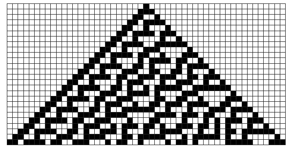
Stephen Wolfram’s A New Kind of Science (NKS) suggests a new way of modelling worldly phenomena. Wolfram postulates that elaborate mathematical models aren’t the only representations of the mechanisms governing the universe; simple patterns may be behind some of the most complex phenomena. In order to illustrate this, he began with cellular automata.

A cellular automaton is a set of colored blocks in a grid that is created stage by stage. The color of each block is determined by a set of simple rules that considers the colors of blocks in a preceding stage.1 Based on just this, cellular automata seem to be fairly simple, but Wolfram illustrated their complexity in rule 30. This cellular automaton, although it follows the simple rule illustrated in Figure 1, produces a pattern that too irregular and complex for even the most sophisticated mathematical and statistical analysis. However, by applying NKS fundamentals, simple rules and permutations of the building blocks pictured can be developed to produce these extremely complex structures or models.2

By studying several cellular automata systems, Wolfram presents two important ideas: complexity can result from simple rules and complex rules do not always produce complex patterns.2

The first point is illustrated by a computer; relying on Boolean logic, the manipulation of combinations of “truths” (1’s) and “falses” (0’s), computers can perform complex computations. And with proper extensions, they can display images, play music, or even simulate entire worlds in video games. The resulting intuition, that complexity results from complexity, is not necessarily true. Wolfram shows again and again that simple rules produce immense randomness and complexity.

There are other natural phenomena that support this theory. The patterns on mollusk shells reflect the patterns generated by cellular automata, suggesting that the shells follow similar simple rules during pattern creation.2 Perhaps other biological complexities are also results of simple rules. Efforts are being made to understand the fundamental theory of physics based on ideas presented in the NKS and Wolfram’s idea might even apply to philosophy. If simple rules can create seemingly irregular complexity, the simple neuronal impulses in the brain might also cause irregular complexities, and this is what we perceive as free will.2

The most brilliant aspect of NKS lies in its underlying premises: a model for reality is not reality itself but only a model, so there can be several different, accurate representations. Our current approach to reality -- using mathematical models to explain the world -- does not have to be the only one. Math can explain the world, but NKS shows that simple rules can also do so. There may be methods and theories that have been overlooked or remain undiscovered that can model our world in better ways.

References

  1. Weisstein, E. W. Cellular Automaton. Wolfram MathWorld, http://mathworld.wolfram.com/cellularautomaton.html (accessed Mar 26, 2016).
  2. Wolfram, S. A New Kind of Science; Wolfram Media: Champaign, IL, 2002

Comment

Dangers of DNA Profiling

Comment

Dangers of DNA Profiling

DNA profiling has radically changed forensics by providing an objectively verifiable method for linking suspects to crimes. Currently, many states collect the DNA of felons in order to ensure that repeat offenders are caught and convicted efficiently.1 Over the past few years, situations in which law enforcement officials can collect DNA from suspects have increased drastically. In 2013, President Obama strongly supported the creation of a national DNA database that included samples from not only people who are convicted, but also those arrested.1 In Maryland v. King (2013), the Supreme Court declared that law enforcement officials are justified in collecting DNA prior to conviction if it aids in solving a criminal case.2 In the years since this decision, the creation of a national DNA database has become a particularly polarizing and contentious issue. Proponents argue that a database would dramatically improve the ability of law enforcement to solve crimes. However, detractors argue that the potential for misuse of genetic information is too great to warrant the creation of such a system.

DNA is popularly referred to as the “blueprint of life” and contains extremely sensitive information such as an individual’s susceptibility to genetic disorders. One of the major arguments against the creation of a national DNA database is that such information could be hacked. Yaniv Erlich, a geneticist at MIT, illustrated this when he used “genome mining” to find the true identities of individuals in a national genome registry. In his study, Erlich obtained genomes from the 1000 Genomes Project, a large database used for scientific research. He then used a computer algorithm to search for specific DNA sequences known as short tandem repeats (STRs) on the Y chromosome of males. These STRs are remarkably invariable from generation to generation. Erlich was able to use the Y chromosome’s STR marker to identify the last names of the individuals to whom the DNA belonged by using easily accessible genealogy sites.3 With just a computer and access to genome data, Erlich could identify personal information in DNA registries. Clearly, the creation of a national DNA database could give rise to widespread privacy concerns. Though there are large fines associated with unauthorized disclosure or acquisition of DNA data, current federal regulations do not technically limit health insurance companies from using genome mining in order to determine life insurance or disability care.4

Hacking of federal databases is not an unreasonable scenario—just this past July, sensitive information including the addresses, health history, and financial history of over 20 million individuals was stolen in a massive cyber-attack.4 That attack uncovered information about every single individual who has attempted to work or has worked in the United States government. A similar abuse of genetic information by third parties is undoubtedly a danger associated with a national DNA database. Despite advances in federal protections such as the Genetic Information Nondiscrimination Act, there are still numerous instances where genetic information regarding disease is used in employment decisions.5

Another potential issue associated with the creation of a DNA database is the notion of genetic essentialism. Genetic essentialism argues that the genes of an individual can predict behavioral outcomes.6 Critics of a national DNA database argue that certain factors—such as the extra Y chromosome—may lead law enforcement officials to suspect certain individuals more than others, which sets up a dangerous precedent.

The notion that chromosomal abnormalities can alter behavioral outcomes has generated numerous studies examining the link between criminality and changes in sex chromosomes—the genes that determine whether an individual is male or female. Normally, females will have two X chromosomes, whereas males have one X chromosome and one Y chromosome. However, in rare cases, males can either have an extra X chromosome (XXY) or an extra Y chromosome (XYY). General literature review suggests that XXY men have feminine characteristics and are substantially less aggressive than XYY or XY men.7 Conversely, studies like Jacobs et al. have suggested that the XYY condition can lead to increased aggression in individuals.8 However, Alice Theilgaard, one of the most prominent researchers on this topic, found that most behavioral characteristics associated with the XYY chromosomal abnormality are controversial.7 Even tests based on objective measures, like testosterone levels, have been inconclusive. Theilgaard argues that the XYY chromosomal abnormality does not cause increased aggression or propensity to commit crimes. Rather, she states that the criminality of XYY individuals might be a socially constructed phenomenon. XYY individuals often have severe acne, lowered intellect, and unusual height. This makes it difficult for people with this condition to “fit in.” As a result of their physical characteristics, XYY individuals might feel ostracized and become antisocial.8 Thus, it is reasonable to conclude that merely having an extra Y chromosome does not predispose someone to be violent; rather a wide variety of social factors play a role.

It is entirely plausible that law enforcement individuals could misinterpret genetic information. For example, they could mistakenly believe that an individual with the XYY condition is more likely to be a suspect for a violent crime. Such an assumption would hinder law enforcement officials from objectively evaluating the evidence involved in a crime and shift the focus to individual characteristics of particular suspects. People in favor of a national DNA database often argue that it would be a great method of solving crimes. Specifically, some officials argue that a database would prevent recidivism (a relapse in criminal behavior) and deter people from committing crimes. However, research done by Dr. Avinash Bhati suggests that the inclusion of DNA in a national registry only seems to reduce recidivism for burglaries and robberies; in other crime categories, recidivism is generally unaffected.9 This suggests that a convict’s knowledge that he/she is in a DNA database is not a true deterrent. The concerns raised by this study should show that databases might not be as effective a crime-fighting tools as proponents suggest.

Both genome mining and genetic essentialism present very real harms associated with the creation of a national DNA database. Having sensitive genetic information in one centralized registry could potentially lead to abuse and discriminatory behaviors by parties that have access to that information. Even if genome databases are strictly regulated, the possibility of that information being hacked still exists. Furthermore, assuming that genetics are the only determinants of behavior could lead to people with genetic abnormalities being suspected of crimes at a higher rate than “normal” individuals. Social factors often shape the way an individual acts; the possibility of law enforcement officials embracing the genetic essentialism approach is another associated harm. In the end, it seems that the negative consequences associated with the creation of a national DNA database outweigh the benefits.

References

  1. Barnes, R. Supreme Court upholds Maryland law, says police may take DNA samples from arrestees. Washington Post, https://www.washingtonpost.com/politics/supreme-court-upholds-maryland-law-says-police-may-take-dna-samples-from-arrestees/2013/06/03/0b619ade-cc5a-11e2-8845-d970ccb04497_story.html (accessed 2015).  
  2. Wolf, R. Supreme Court OKs DNA swab of people under arrest. USA Today, http://www.usatoday.com/story/news/politics/2013/06/03/supreme-court-dna-cheek-swab-rape-unsolved-crimes/2116453/ (accessed 2015).
  3. Ferguson, W. A Hacked Database Prompts Debate about Genetic Privacy. Scientific American, http://www.scientificamerican.com/article/a-hacked-database-prompts/ (accessed 2015).
  4. Davis, J. Hacking of Government Computers Exposed 21.5 Million People. The New York Times, http://www.nytimes.com/2015/07/10/us/office-of-personnel-management-hackers-got-data-of-millions.html?_r=0 (accessed 2015).
  5. Berson, S. Debating DNA Collection. National Institute of Justice, http://www.nij.gov/journals/264/pages/debating-dna.aspx (accessed 2015).
  6. Coming to Terms with Genetic Information. Australian Law Reform Commission , http://www.alrc.gov.au/publications/3-coming-terms-genetic-information/dangers-‘genetic-essentialism’ (accessed 2015).
  7. Are XYY males more prone to aggressive behavior than XY males? Science Clarified, http://www.scienceclarified.com/dispute/vol-1/are-xyy-males-more-prone-to-aggressive-behavior-than-xy-males.html (accessed 2015).
  8. Dar-Nimrod, I.; Heine, S. Genetic Essentialism: On the Deceptive Determinism of DNA. Psychological Bulletin, http://www.ncbi.nlm.nih.gov/pmc/articles/pmc3394457/ (accessed 2015).Are XYY males more prone to aggressive behavior than XY males? Science Clarified.
  9. Bhati, A. Quantifying The Specific Deterrent Effects of DNA Databases. PsycEXTRADataset. 2011. https://www.ncjrs.gov/app/publications/abstract.aspx?id=258313 (accessed May 2015).

Comment