Viewing entries in
Biological Sciences

It’s in the Genes: Advances in Genome-Editing Tools

Comment

It’s in the Genes: Advances in Genome-Editing Tools

by Esha Ghai

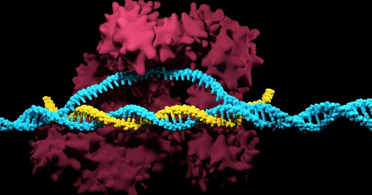
We have long been told that our DNA is unchangeable, often encouraging us to blame our flaws and accredit our talents to genetics. However, the introduction of genome-editing gives scientists the ability to alter genetic material and effectively change the basis of an organism. Genetic material can now be added, removed, or mutated at any specified location in an organism, allowing for cures to genetically inherited diseases and a wide range of illnesses. A recently discovered microbial ribonucleoprotein with such genome-editing capabilities is the CRISPR/Cas9 system. 

CRISPR/Cas9, short for “clustered regularly interspaced short palindromic repeats and CRISPR-associated protein 9”, was naturally developed by bacteria as a defense mechanism against invading viruses. As a part of this mechanism, bacteria confiscate fragments of DNA from invading viruses, and use the fragments to construct CRISPR arrays [1]. CRISPR arrays give the bacteria the ability to recognize identical or similar viruses, so if a similar virus attacks again, the bacteria produces RNA segments from the CRISPR array to specifically attack the virus’s DNA [1]. Cas9, an enzyme, is then used to cut apart the DNA, essentially killing the virus [1]. Scientists in the medical community are now adapting this gene-manipulation system to be used in cells in other organisms, including humans. The genome-editing tool has unlocked the potential to combat human genetic diseases previously deemed untreatable. A more cost-efficient, advanced, and precise method of gene-editing, CRISPR/Cas9 is revolutionizing the face of medicine as we know it [1].

Fascinated by the properties of CRISPR/Cas9 and microbes, Dr. Xue Gao, a professor of Chemical and Biomolecular Engineering at Rice University, currently investigates the applications of genome-editing and microbe-based natural product discovery. Her research “lies at the interface of chemical biology and biomolecular engineering with a primary focus on small- and macro-molecule discovery and their applications to human health, agriculture, and energy.” Dr. Gao’s contribution to the development of Statins, a pharmaceutical drug with lipid-lowering capabilities for those at risk of cardiovascular disease, piqued her interest in protein engineering. The frequent use of Statins in the medical society motivated Dr. Gao to continue synthesizing molecules to combat diseases. 

Her research also encompasses the use of enzyme-catalyzation (increase in the rate of a process due to an enzyme) for anticancer and antibiotic drug synthesis. Enzymes are powerful biological catalysts, accelerating chemical reactions by assisting in the formation of a transition state between the starting reagent and end product. Enzymes are also highly specific in which reactions it catalyzes, usually catalyzing only a single chemical reaction (or reactions very similar in nature), limiting side reactions and the ultimate formation of wasteful by-products [3]. According to Dr. Gao, whereas traditional chemical methods of synthesizing compounds require environmentally toxic solvents and harsh conditions, enzymes work well in moderate environments and are capable of producing a higher yield of 99%. As enzymes are proteins, extreme temperatures and pH levels result in denaturation, rendering the enzyme inactive. Therefore, enzyme catalyzation ability is enhanced at moderate conditions. In biological systems, a majority of processes utilize enzymes, from the transfer of CO2 in the body through the enzyme carbonic anhydrase, to breaking down sugars through the enzyme amylase. The development of enzymes to catalyze complex chemical reactions, called directed evolution, is a major focus of the Gao Laboratory. 

Dr. Gao ascribes “her love [for] exploring what has already been designed by nature” to the sheer intelligence of microbes and their methods of defense against viruses. “Microbes are super smart. They create protein encodings themselves to fight different types of environments.”  CRISPR/Cas9, another prominent example utilizing protein encodings and enzyme catalysis, has also attracted Dr. Gao’s interest. CRISPRs are specialized protein encodings, while Cas9 is an enzyme capable of cutting apart genetic material. Recently, Dr. Gao utilized the genome-editing capabilities of CRISPR/Cas9 to treat autosomal-dominant hearing loss. Autosomal-dominant diseases are inherited from an abnormal gene from one parent, even when the corresponding gene from the other parent is normal. Dr. Gao’s findings suggested that a targeted delivery of gene-disrupting agents such as CRISPR/Cas9 in vivo (within a living organism) can potentially treat such cases of hearing loss [2].

Dr. Gao’s research group is planning to do further work in CRISPR/Cas9 applications for a variety of human genetic diseases, and hopes to make breakthroughs in research through accommodating advanced computational tools and developing DNA sequencing technology (technology that determines the sequence of nucleotide bases). Dr. Gao envisions the next stage of her group’s research to be developing new enzyme-catalyzed reactions in the energy sector, particularly the production of biofuels, and combating food contamination in the agricultural industry. 

Dr. Gao’s current and future research aspirations in the field of human genetics and the energy sector serve as an inspiration for aspiring female scientists globally. She hopes that young females will follow their passion in STEM, and envisions a future where female scientists are not in the minority. Because of Dr. Gao and her vision, the Gao Laboratory is not only an embodiment of cutting-edge innovation in the future of enzyme-catalyzed reactions, but also the inspirational potential of female scientists. 

Works Cited

[1] What are genome editing and CRISPR-Cas9? - Genetics Home Reference - NIH. https://ghr.nlm.nih.gov/primer/genomicresearch/genomeediting (accessed Nov 8, 2019).

[2] Gao, X., Tao, Y., Lamas, V. et al. Nature. [Online] 2018, 553, 217-221. https://www.nature.com/articles/nature25164 (accessed Nov 8, 2019).
[3] Berg, J.; Tymoczco, John; etc. Biochemistry, 5th; W.H Freeman & Co: New York, 2002; 303-341.

Comment

Comment

Membrane Fusion And The Secret to Muscular Degeneration

by Lam Nguyen

In the human body, there are about 37 trillion cells. Each cell is made up of organelles that are busy creating proteins, digesting nutrients, and transporting different molecules or other organelles around. In order to facilitate the diverse functions different organelles assume, the plasma membrane, a barrier made up of proteins and lipids, helps compartmentalize different metabolic reactions to specific organelles and regions of the cell. Plasma membranes also play a critical role in the transportation of nutrients and signals between organelles through a process called fusion. A major topic within plasma membrane research, membrane fusion is a process in which two separate lipid membranes fuse into one single layer [1]. This process is essential in the creation of many organelles, such as the ER, a particular organelle that has fascinated Dr. James McNew, professor of biosciences, ever since he first came to Rice University.

Dr. McNew received his Bachelor’s of Science in Biochemistry from Texas A&M University, his Ph.D. in Pharmacology from UT Southwestern,  and he completed his postdoctoral fellowship at the Memorial Sloan-Kettering Cancer Center. His interest in cellular biology and membrane fusion started with his graduate work while researching how the peroxisome, an organelle responsible for various metabolic processes such as detoxification and cellular signaling, worked. This particular lab experience helped him realize that he had a passion for understanding basic molecular biology and “how different cellular components moved around the cell.” His burgeoning interest in basic cell biology followed him to his postdoctoral fellowship where his research centered around how proteins were moved, targeted, and carried to the Golgi apparatus, an organelle that processes and sends out newly synthesized molecules. Dr. McNew’s passion for cell biology and membrane fusion  has defined much of the work he has done here at Rice. His original research pertains to understanding membrane fusion in the endoplasmic reticulum (ER). The ER is a network of membranous tubules that is involved in protein and lipid synthesis1. Dr. McNew and Dr. Michael Stern, a fly geneticist and professor at Rice, observed how these tubules came together, but “the proteins involved were unclear.” They then hypothesized that the protein elastin, a protein present in connective tissue, played a significant role in ER membrane fusion. The two were able to confirm that elastin helps ER tubules come together, and they characterized the protein by studying it both in vivo (in a test tube) and back in fruit flies.

Dr. McNew also describes himself as a “basic cell biologist” and that his work is generally “far removed from something medicinal or therapeutic.” Work by clinicians on the elastin protein, however, has confirmed that elastin plays a role in physiological processes in humans. In fact, defects in the gene encoding for elastin play a role in hereditary splasic paraplegia (HSP), a group of genetic diseases where weakness and stiffness in the muscles gradually lead to muscular degeneration overtime.   This discovery has allowed Dr. McNew to connect his basic biology research to clinical applications. Alongside characterizing the mechanism of elastin in ER membrane fusion, Dr. McNew has also been deepening his understanding of how mutations in this protein can cause human diseases. 

Trying to understand how disruptions in the elastin and ER membrane fusion could cause the degeneration of muscles seen in HSP, Dr. McNew’s lab discovered that the loss of elastin function results in neurodegeneration which eventually leads to the deterioration of muscles. This explains how “people who have HSP lose motor control and their ability to walk.” Between adjacent neurons, small signaling molecules called neurotransmitters are transported  to send the electrical signal from one neuron to another. Dr. McNew observed that defects in elastin seen in HSP patients are somehow connected to the lack of neurotransmission. The lab was initially puzzled because they saw signs of muscle degeneration but not neurodegeneration. “Even though nerve damage is a known cause of muscle atrophy, conventional wisdom is that the nerve cells die first," Dr. McNew explains.  Consequently, Dr. McNew and his lab have begun investigating the cellular communication involved in this seeming paradox by learning about the other signalling molecules involved, specifically the torr kinase protein [2]. The McNew lab has been researching how the lack of neurotransmission is signalled by the torr kinase in order to provoke cell death in muscular tissue. 

Dr. McNew plans on building on the vast amount of data that his lab has obtained by further exploring other types of proteins that work with elastin and torr. Understanding these biochemical interactions will allow scientists to have a better picture of how elastin defects, lack of neurotransmission, and muscular degeneration are all connected to each other. The opportunity to discover new conclusions and to be on the forefront of scientific innovations is what often inspires undergraduates to get involved in research. When asked about if he has any advice for aspiring undergraduate researchers, he says that one has to be “okay with failure.” “It is very exciting and engaging to do something for the very first time. The feeling that you are the first person to do something is very addictive and keeps you going!” exclaims Dr. McNew. It is this excitement that will continue to inspire Dr. McNew in furthering his understanding of our cells and how they work.


Works Cited

(1) Daumke, O.; Praefcke, G. Structural Insights Into Membrane Fusion At The Endoplasmic Reticulum. Proceedings of the National Academy of Sciences 2011, 108 (6), 2175-2176.

(2) Loewith, R.; Hall, M. Target Of Rapamycin (TOR) In Nutrient Signaling And Growth Control. Genetics 2011, 189 (4), 1177-1201.


Comment

Comment

The Machines within Our Cells: Rewiring Genetic Circuits

by Gulnihal Tomur

Many people regard biology as simply trying to solve the mysteries of nature through observation.  A newly emerging field called synthetic biology bridges the gap between engineering and biosciences through reconstructing  already existing biological systems or fabricating entirely new ones by using artificial DNA synthesis technology. As synthetic biologists construct and insert manufactured genes in cells, they have two main goals in mind: either turn the cells into drug or biofuel factories or gain a better understanding of the roles different biological components play in naturally existing biological system. Synthetic biology allows scientists to reconstruct existing biological systems on a molecular level.

When asked to describe synthetic biology, Dr. Caleb Bashor of the BioScience Research Collaborative at Rice alludes to a famous quote from physicist Richard Feynman: “What I cannot create, I do not understand.” Dr. Bashor and his team attempt to rebuild intricate biological pathways to better comprehend their underlying principles. He refers to cells as micro-level machines — and “just like any machine, the cells have wiring inside them in the form of DNA and proteins.” Accordingly, Dr. Bashor’s approaches cellular systems from an engineering standpoint as he reconstructs these systems just like how an electrical engineer would rewire a machine to figure out how it works. “I am partly an engineer and partly a scientist, trying to understand how cells are organized so I can rewire them,” Dr. Bashor explained. 

Yet, Dr. Bashor did not start his scientific journey as a synthetic biologist; instead, he first occupied himself with biochemistry as an undergrad at Reed College. He was especially drawn to thinking about biology from the protein structure scale. At UCSF, he pursued the study of biological structures as a grad student from a perspective rooted in biophysics. Dr. Bashor got involved in synthetic biology at its early stages through a protein structure and function lab at UCSF, where they put different protein domains together to change the function of the protein as it was expressed in the cell. Dr. Bashor shares how they were essentially engaged in synthetic biology, yet did not know how broad the scope of their research was: “Eventually, people came along and told us ‘You guys are doing synthetic biology.’ And our reaction was ‘What’s that?’” As Dr. Bashor immersed himself more and more into this field, he dedicated his postdoc research at MIT to engineering transcriptional networks in eukaryotic cells. His current research at BioScience Research Collaborative encapsulates an even more ambitious approach as he and his team establish “synthetic regulatory circuits to reprogram the behavior of human cells.” Dr. Bashor and his team currently tackle mammalian regulatory circuits with potential applications that range from biosensing to cell-based therapy.

In order to fully understand Dr. Bashor’s work and its implications, we must first look into a natural gene circuit. Both eukaryotes and prokaryotes carry multiple genes in their genome, some of which get turned on and off depending on the external and internal signals cells receive. Since most proteins have complex structures, usually multiple genes are clustered together in a transcription unit. This then codes for the subunits of a single protein. The expression of these  genes are all dependent on a single promoter for their expression; the promoter serves as the site where RNA polymerase binds to DNA to initiate the transcription of the information embedded in DNA to mRNA. The so-called on-off switch of such transcription units is a specific DNA segment called an operator. Located either within or after the promoter, the operator is responsible for regulating when RNA polymerase can bind to DNA and start the transcription of the gene cluster. The promoter, the operator, and the clustered genes make up genetic circuits known as operons. 

When another protein called a repressor, which is the product of a regulatory gene separate from the operon’s gene cluster, is present in its functional state, it binds to the operator, thus physically blocking RNA polymerase from binding to the promoter. Internal and external factors, such as the presence of a specific molecule, influence the activity of the repressor by changing its 3D shape, which allows cells to switch on and off specific gene clusters based on environmental cues. Synthetic biologists take advantage of this knowledge as they introduce molecules in the cell which may turn on or off a repressor, add artificially designed circuitry into the existing systems, or isolate certain parts of a naturally occurring gene circuit through genetic modification. 

Mirroring and reconstructing such systems, however, still proves to be a real challenge despite the advancement in synthetic biology. One common problem scientists deal with is the instability of artificial gene circuits. These artificial circuits can break the natural balance of the metabolic systems of the cell. As the artificial gene circuits use the energy which would otherwise be used for growth and division, they hinder the cells from maximizing their fitness. Thus, any mutant cell in the cluster which did not accept the circuit outcompete the synthetically engineered cells. “The circuit becomes unstable evolutionary; the cells spit out the circuit over time in order to grow faster,” explains Dr. Bashor. Intrigued by this evolutionary process, he put his mind to engineering a circuit that doesn’t get spat out. He realized that he needed a device that would help him and his team monitor the evolutionary process of cells with artificial circuits in them. The result was eVOLVER — it enabled them to work with many circuits at a time. As an automated multi-culture platform, eVOLVER can control multiple parameters at once. It shows the cell growth and detects any disparities which may signal a mutant culture that spat out the circuit put into the system. “We can grow [the cells], watch the circuits break, and see where they recover their fitness as a result of the circuit breaking,” tells us Dr. Bashor. Ultimately, the device operates as a tool for being able to build circuits and then test their stability, which is a crucial factor if the circuit is desired to be stable for a long time so that, for instance, it can be used for the production of a chemical.

Another challenge scientists face presents itself in the complexity of multi-gene systems. Pleitropic genes, which influence two or more seemingly unrelated phenotypic traits, and gene clusters regulated by common factors hinder scientists who aim to understand the function of individual parts in such systems. Therefore, synthetic biologists turn to a technique called refactoring. “Refactoring is a term borrowed from computer science,” tells us Dr. Bashor, “somebody gives you a code that is really hard to work with, so you rewrite in a way that it is easier to change the parts individually and to see what you’re changing.” In a similar fashion, synthetic biologists “physically abstract genes away from their native genomic content.” (1) The new content in which scientists place the gene in question tends to be a simple plasmid. Not only does this new medium offer easier control over the effects and outcomes of the gene due to its simplicity, but it also allows scientists to quickly modify the new genomic content since putting a new piece of DNA in a plasmid is a relatively easy process. By eliminating the interference caused by the natural genomic context, synthetic biologists observe the role of specific genes in the whole system. Yet, Dr. Bashor warns us not to see interference as a sheer burden on the system. “You may make the regulation very simple, but then you lose something in the behavior. That’s a clue that the regulatory forces that are associated with that context are important.” In the end, he sees refactoring, and synthetic biology in general, as a trial-and-error process — synthetic biologists simplify and modify and rewire the systems they are curious about over and over again to master the role of individual parts, which then allow them to see the bigger picture more clearly.

There are many other obstacles in the synthetic biology scene scientists are still trying to overcome today. The inability to insert big chunks of DNA into cells, for example, is hindering scientists from taking full advantage of synthetic biology in cell therapy. Yet, Dr. Bashor sees the intersection of synthetic biology and cell therapy as one of the most promising developments to look out for in the future. As the trial-and-error process that is synthetic biology continues to ignite curiosity in current and future scientists, the field will offer more and more to the scientific community. From advancements in agriculture to biofuel production, the manipulation of biological systems has already enhanced our lives in ways we may not even realize, and it continues to help us finesse biological systems in ways we desire.

Sources

Synthetic Biology Explained. https://archive.bio.org/articles/synthetic-biology-explained (accessed Jan 20, 2020).

The Bashor Lab at Rice. http://bashorlab.rice.edu/index_large.html (accessed Jan 20, 2020).

Comment

Comment

A Stressful Situation

by  Saksham Vashistha

Affecting nearly 80% of Americans, stress has become an integral part of our communities. However, while stress can affect our mental health, cells, too, experience physical stresses. However, while stress can affect our mood, physical stresses affect our bodies at the cellular level. Getting to the bottom of how cells respond to stresses is a useful tool in a wide variety of fields. Dr. Mike Gustin, a professor of biological sciences at Rice University, aims to understand stress responses in cells and their role in the treatment of cancer.

Dr. Michael Gustin’s passion for cellular biology developed during his time as a graduate student. While working in the Ching Kung lab at the University of Wisconsin-Madison, then-physiologist Dr. Gustin and his team were the first to discover that ion channels, special proteins that span the cellular membrane and help transport charged particles into and out of the cell, exist in yeast and bacteria. Now at Rice University, Dr. Gustin has made the transition to genetics, focusing on the impact of stress on cellular behaviors. His most recent works include collaborations with researchers across the Houston area, namely Dr. Erik Cressman, a chemist at MD Anderson. 

Dr. Gustin and Dr. Cressman worked together on an NIH funded project that investigates ways to treat liver cancer. Unfortunately, as Dr. Gustin points out, “for liver cancer, there is no good chemotherapy.” Oftentimes, cancerous tumors in the liver are inoperable and alternative treatments are required. However, Gustin and his team have found that “chemistry-driven exothermic reactions (heat-generating reactions) can ablate (remove) tissues in an increasingly controlled way.” There are a variety of different reactions that can be used to ablate cells. Dr. Gustin’s group decided to use a reaction between an acid and a base. By mixing the two solutions on the cell’s surface and allowing them to react, heat and a high solute concentration (osmolarity) are produced. This extreme heat combined with the high osmolarity is toxic to the cell and kills it.

As Dr. Gustin explains, “there is an ablation zone [on the cell’s surface] where the temperature is high enough to kill cells and there is a combination of not just heat, but also high osmolarity because of the salt that accumulates there.” Gustin’s group is interested in the peri-ablation zone, the area just around the region of high osmolarity.  It was hypothesized that the applied cellular stresses are followed by some types of cellular defense mechanisms or signaling pathways. These signaling pathways serve to protect the cell from physiological stresses, helping it survive the harsh ablation conditions. Understanding how these pathways work is key for ablation treatment in cancer patients. The goal is, as Dr. Gustin says, “to inhibit those pathways in a controlled way [to] control how much ablation actually happens.” Thus, by inhibiting certain cellular defense mechanisms, ablation in cancer cells can be modulated and used precisely during cancer treatment. 

One of the interesting finds during their investigation was that sometimes the stress applied to the cells prevented the cells from even responding. Upon further investigation, it was found that some of the genes that are activated for the stress response actually inhibited other stress responses. Dr. Gustin’s team attempted to understand these stress responses at the molecular level. By using computer modeling of known proteins that were involved in the stress response pathways, they were able to identify how the structures of the proteins were changed by the cellular stresses. Gustin’s team wanted to understand “what happens to the fluid around a protein and what happens when stresses combine around that protein.” Understanding what happens to the fluid around the protein can indicate what environmental changes the protein is experiencing. The alteration of the fluid condition can lead to altered protein structures. Changes in the molecular structure of proteins affects the cell’s ability to react properly to stress. This is because the structure of the protein is what determines its function. If the structure is altered in any way, the protein wouldn’t be able to carry out its function correctly. This molecular modeling approach can help scientists understand not only at the cellular but also the tissue and organ level of how stress responses operate in a mammalian body. Furthermore, ablation in tumor cells can be controlled via inhibition of the genes and subsequent proteins involved in the cellular stress response. This control over the cell’s genes can help make ablation more effective for liver cancer patients with inoperable tumors. 

Dr. Gustin’s investigation into harnessing the power of cellular stresses has the potential to change how physicians approach the treatment of cancer. By identifying mechanisms by which cancer cells resist ablation, Gustin’s work provides new insight into how physicians can directly alter cellular defense mechanisms to make them more vulnerable to treatment. While understanding the influences of stress on cells has made significant contributions in medicine, Gustin’s work can also have applications in other fields. Dr. Gustin has his eyes set on something greater. “I am really curious about … what happens during coral reef bleaching”, Dr. Gustin says with a glint in his eyes. Coral reef bleaching is a direct result of stresses (changing water temperatures) on the cells of the organism, causing them to lose the algae living in them. The algae are responsible for providing the majority of the coral’s energy, without them the coral starve and die.  Gustin hopes to use his knowledge of cellular stresses to “genetically engineer algae that stay in the coral” reagrdless of the water temperature in an effort to save an inumerable amount of coral around the world. Such an effort would help lead the effort to rebuild important coral reef ecosystems globally.

[1] Saad, L. Eight in 10 Americans Afflicted by Stress. https://news.gallup.com/poll/224336/eight-americans-afflicted-stress.aspx

[2] Fuentes, D.; Muñoz, N. M.; Guo, C.; Polak, U.; Minhaj, A. A.; Allen, W. J.; Gustin, M. C.; Cressman, E. N. K. A Molecular Dynamics Approach towards Evaluating Osmotic and Thermal Stress in the Extracellular Environment. International Journal of Hyperthermia2018, 35 (1), 559–567.

Comment

Comment

ATAC: A Brain Therapy of the Future?

by Sophianne Loh

Rice University students and faculty are said to possess “unconventional wisdom.” We aren’t afraid to dream of solutions that seem impossible, push past limited frameworks of thought, and leap into uncharted territory. This ideal is perhaps most apparent in the neuroengineering research done at Rice. For years, neurological conditions like Alzheimer’s or epilepsy have been treated in much the same way: surgical procedures, injections, and electrical stimulation of the brain. [1] These treatments aim to “fix” dysfunctional neural circuits, which are often the origin of these neurological and psychological disorders. Unfortunately, not only are these options invasive, but they fail to target specific dysfunctional neurons, leading to adverse effects on the surrounding healthy brain tissues. 

As a result, researchers around the world have been hard at work creating smaller implants and less invasive electrodes on the scales of millimeters or even micrometers. [2] For example, researchers at the Korea Advanced Institute of Science and Technology recently designed a quarter-sized wireless implant that successfully modulated mouse brain neurons. [3] However, continuing to travel down the same path of surgical implants eventually leads to the same major issues: unwanted invasiveness, limited specificity of targets, and, most prominently, obtaining sufficient funding. As Dr. Jerzy Szablowski of the Rice Bioengineering Department points out, “If you look at the amount of funding we have for brain disorders - for example, from National Institutes of Health - you'll find out that the amount of money we spend on research that aims to treat brain disorder is very low compared to the burden that they cause” In fact, Dr. Szablowski notes the cost of creating one new drug for each brain disorder can be up to $1 billion. In comparison, the National Institute of Mental Health dedicated only $2.1 billion in 2021 to research on brain disorder treatments. [4] With over 1,000 neurological disorders affecting over 100 million individuals in America, there is an ever-growing need for less costly and more versatile treatments. [5]

In true Rice fashion, Dr. Szablowski approached this need by traveling down a different route. He wanted to create a one-stop, noninvasive, affordable treatment for multiple neurological disorders. In other words, what if a single technology could treat a variety of  neurological disorders with elegant simplicity? Dr. Szablowski has found an answer in acoustically targeted chemogenetics, or ATAC. Developed by Szablowski and advanced by his researchers in the Laboratory for Noninvasive Neuroengineering, ATAC is the first-ever fully noninvasive technique to control neurons with spatial, cell-type, and temporal precision. When applied to the treatment of neurological disorders, ATAC could treat a variety of disorders, as long as they rely on abnormal neuronal activity.

Despite the simplicity of the resulting treatment path, ATAC is a complex, multi-step process. It starts with the blood-brain barrier (BBB), a collection of different types of cells tightly joined together that line the blood vessels of the brain. The BBB serves to block certain pathogens or other harmful molecules in the bloodstream from entering the brain. However, this also means that the BBB won’t normally allow the passage of therapeutic molecules into target areas in the brain. [6] This is where the Szablowski lab turned to FUS-BBBO, or focused ultrasound blood-brain barrier opening, in their development of the ATAC method.

FUS-BBBO is a technique that had been well-researched prior to Dr. Szablowski’s development of ATAC. This ability to build upon a foundation of past research while pioneering a new treatment was important to Dr. Szablowski. He explains, “I wanted to find a method that is well-validated and something that would be safe for human use, and the fastest way to do that is to just look at what is available.” FUS-BBBO (Fig. 1) first involves the injection of microbubbles (typically a lipid or protein shell containing perfluoropropane or sulfur hexafluoride gas) into the desired blood vessels. Immediately after, short, low-pressure ultrasound wave pulses are applied to the BBB target site. This causes the microbubbles in the blood vessels to oscillate, a phenomenon termed stable cavitation. The oscillations apply pressure to the surrounding fluid, which opens the tight junctions between the cells of the BBB temporarily and reversibly. As a result, therapeutic molecules in the bloodstream can go past the BBB to reach neurons in the brain. [7]


Figure 1. (A) Schematic depiction of FUS-BBBO procedure. (B) MRI-guided FUS-BBBO procedure. [6]

Where Dr. Szablowski departs completely from previously known procedures is by combining FUS-BBBO with AAVs, or adeno-associated viruses, that encode engineered protein receptors that can control neurons. An AAV is known as a “vector” for gene therapy. This means scientists can insert desired DNA into the AAV allowing it to deliver and express these genes to certain cells in the body. [8] In ATAC, FUS-BBBO first opens specific areas in the BBB for AAV to deliver its genes. In this way, FUS-BBBO essentially “marks” the specific neural circuits to be treated, contributing to the spatial specificity of the ATAC treatment. The AAV vectors contain DNA that codes for engineered proteins called DREADDs, a type of cell receptor. These cell receptors allow a cell to respond to designer drugs, which are drugs created in the lab, that can get into -  but do not activate naturally-occurring receptors in - the brain. When the AAV vector delivers the DREADD genes to the specific neurons marked by the FUS-BBBO procedure, the expression of the DREADDs allows the neurons to be activated by designer drugs. The designer drug used in the ATAC procedure is called clozapine-N-oxide (CNO). CNO has the ability to excite or inhibit certain neurons that express DREADDs, allowing for targeted treatment of neurological disorders. Furthermore, it can be administered in a non-invasive way to patients (i.e., as a pill). [9] It is the combination of FUS-BBBO, gene therapy through the AAV vectors, and CNO that makes ATAC so effective at targeting specific neurons. (Fig. 2)

Figure 2. Depiction of the ATAC procedure. [1]

The Szablowski lab chose to test ATAC by observing its effect on the hippocampus of rodents. Dr. Szablowski describes the hippocampus as “a very relevant brain region for many disorders - epilepsy being one of them. ” So, if ATAC is effective on the hippocampus, then it has the potential to treat a multitude of disorders, ranging from epilepsy to PTSD. Specifically, initial tests focused on how ATAC could modulate memory formation, providing a possible treatment for anxiety disorders. To test the efficacy of the treatment, rodents were exposed to a fear-inducing stimulus in a unique experimental setting. Next, both the rodents who had received AAV and DREADDs and those in a control group received either CNO or saline solution. Twenty-four hours after initial exposure to the stimulus, the rodents were again placed in the same experimental setting to test their fear recall.

The results suggest that ATAC applied in the hippocampus successfully reduced memory formation. In a common memory task, ATAC rodents that received CNO showed less than half of the response than rodents in the control group. Furthermore, the results showed ATAC only targeted memory formation and had no effect on exploratory behavior and sensation of stimuli, confirming the high specificity of the treatment. What does this all mean for us? Well, if ATAC becomes clinically relevant, it could enable a minimally invasive, highly effective way of treating neurological or psychiatric disorders including Parkinson’s disease or epilepsy. As Dr. Szablowski describes, “any [neuropsychiatric disorder] that has the defined activity of the [neural] circuit that needs to be modified” could potentially be treated with ATAC. 

In practice, Dr. Szablowski envisions that ATAC would first involve an outpatient procedure that opens the BBB and delivers the gene therapy. After waiting about three weeks, the patient would simply take pills regularly to deliver the CNO to the “painted” areas of the brain. There would no longer be a need for implanted devices and multiple intracranial injections. It’s a streamlined, powerful form of treatment that can greatly simplify what a patient must go through while seeking relief from neurological disorders. However, ATAC still faces a long journey toward reaching clinical relevance. Like all biomedical research endeavors, ATAC must overcome many obstacles. For example, Dr. Szablowski recognizes that ATAC would be a very expensive procedure with current technology and techniques. So, his lab is currently developing accessory technology for ATAC, such as “site-specific therapeutics,” a technique that can be used before AAV vectors to temporarily modulate a specific site of the brain. This would allow medical practitioners to test whether or not their chosen target in the brain for ATAC is indeed correct, based on the disorder and the individual. Site-specific therapeutics would help prevent the unfavorable situation in which expensive, irreversible, and potentially risky gene therapy has been applied to the incorrect site in the brain during ATAC. Once ATAC is ready for  clinical trials, they’ll start testing this combination of site-specific therapeutics and ATAC for the treatment of Parkinson’s disease.

With careful planning and creative thinking, the Szablowski lab has created a minimally invasive neurological disorder treatment that maximizes effectiveness and simplifies procedures. While ATAC faces and will continue to face obstacles on its path toward implementation in the medical field, this unique solution brings us closer to a future of accessible and optimized biological treatments for complicated disorders.


Works Cited

[1] Szablowski, J. O.; Lee-Gosselin, A.; Lue, B.; Malounda, D.; Shapiro, M. G. Acoustically Targeted Chemogenetics for the Non-Invasive Control of Neural Circuits. Nature Biomedical Engineering 2018, 2 (7), 475–484. 

[2] Kozai, T. D. Y. The History and Horizons of Microscale Neural Interfaces. Micromachines 2018, 9 (9), 445. 

[3] Kim, C. Y.; Ku, M. J.; Qazi, R.; Nam, H. J.; Park, J. W.; Nam, K. S.; Oh, S.; Kang, I.; Jang, J.-H.; Kim, W. Y.; Kim, J.-H.; Jeong, J.-W. Soft Subdermal Implant Capable of Wireless Battery Charging and Programmable Controls for Applications in Optogenetics. Nature Communications 2021, 12 (1). 

[4] 2021 Autumn Inside NIMH. https://www.nimh.nih.gov/research/research-funded-by-nimh/inside-nimh/2021-autumn-inside-nimh (accessed Apr 6, 2022). 

[5] Gooch, C. L.; Pracht, E.; Borenstein, A. R. The Burden of Neurological Disease in the United States: A Summary Report and Call to Action. Annals of Neurology 2017, 81 (4), 479–484. 

[6] Daneman, R.; Prat, A. The Blood–Brain Barrier. Cold Spring Harbor Perspectives in Biology 2015, 7 (1). 

[7] Wang, J. B.; Di Ianni, T.; Vyas, D. B.; Huang, Z.; Park, S.; Hosseini-Nassab, N.; Aryal, M.; Airan, R. D. Focused Ultrasound for Noninvasive, Focal Pharmacologic Neurointervention. Frontiers in Neuroscience 2020, 14.

[8] Naso, M. F.; Tomkowicz, B.; Perry, W. L.; Strohl, W. R. Adeno-Associated Virus (AAV) as a Vector for Gene Therapy. BioDrugs 2017, 31 (4), 317–334.  

[9] Burnett, C. J.; Krashes, M. J. Resolving Behavioral Output via Chemogenetic Designer Receptors Exclusively Activated by Designer Drugs. The Journal of Neuroscience2016, 36 (36), 9268–9282.

Comment

Comment

Evaluating the effectiveness of CQ and/or HCQ against SARS-CoV-2

by Puneetha Goli

Abstract

Severe acute respiratory syndrome coronavirus 2 (SARS-CoV-2) is the virus that causes the coronavirus disease (COVID-19). Patients exhibit a spectrum of severity in their symptoms ranging from asymptomatic to breathing difficulties[1]. The purpose of this article is to evaluate the effectiveness of chloroquine (CQ) and its less toxic metabolite hydroxychloroquine (HCQ) as a viable treatment for SARS -CoV-2 through analyzing data from available research studies. In one of the early breakthrough studies published in February 2020, Gao et al. found that a low-micromolar concentration of CQ blocked COVID-19 infection in early in vitro studies and improved lung imaging, indicating a potential correlation between CQ administration and a decrease in the spread of COVID-19[2]. However, more recent studies, including Skipper et al., since then have indicated that there are no significant differences with the administration of the agents, and some even caution against the use of them because of potential long-term side effects [3,4]. The general consensus among the scientific community, however, remains that the use of agents must be rigorously considered on a case-by-case basis and with caution in research or clinical trial settings[4]. Determining the effectiveness of CQ/HCQ will aid in preventing the spread of misinformation. 


Introduction 

Responsible for taking the lives of 2,244,713 people worldwide (as of February 3, 2021), the World Health Organization (WHO) classified COVID-19 as a pandemic in March 2020 (5,6). SARS-CoV-2, the virus responsible for COVID-19, is only one of seven identified human coronaviruses and is primarily transmitted from person-to-person through direct contact, saliva, and airborne droplets. It is considered “novel” because it is newly identified and is different from the other coronaviruses that have been linked with causing mild illnesses, such as the common cold [7]. Because of its novel nature, there is much that is unknown about the virus and the disease, and unfortunately misinformation regarding treatments has been spread, specifically the effectiveness of chloroquine (CQ)/ hydroxychloroquine (HCQ) as a treatment [7]. 

CQ and HCQ are antimalarial treatments, and interest in these drugs as possible treatments for COVID-19 was spurred in part from the drugs’ history of in vitro activity against viruses such as influenza. HCQ’s role in modulating the immune system through treating other autoimmune diseases, including systemic lupus erythematosus and rheumatoid arthritis, could have also played a role [4,7].  

In April 2020, nearly 55 countries, including the US, Mauritius, and Seychelles, were in the process of receiving HCQ from India [8]. Country leaders, including Brazil’s President Jair Bolsonaro and United States’s President Donald Trump, showed support for the drug, some even claiming to have taken it [9,10]. 

In the months since, however, scientists and national agencies have discouraged the use of CQ and HCQ against treatment for COVID-19, claiming that its effectiveness hasn’t been proven yet. Evaluating the research surrounding CQ and HCQ’s effectiveness is critical in order to address misconceptions and misinformation that is being spread as well as to evaluate its role as a potential treatment.

This paper will be reviewing two studies presenting contradictory evidence regarding the effectiveness of CQ/HCQ. In the first study published on March 16, 2020, Gao et al. examines the effectiveness of CQ in the early stages of the COVID-19 pandemic. Later, a study published on October 20, 20202 by Skipper et al., provides a more comprehensive comparison documenting HCQ’s utility.

Materials and Methods 

Gao et al., one of the early breakthrough studies which found favorable results in treating pneumonia associated with COVID-19 with CQ/HCQ, first conducted in vitro studies in which CQ was tested against the COVID-19 infection [2]. After a determination of a half-maximal effective concentration (EC50) and a half-cytotoxic concentration (CC50), multiple clinical trials were then conducted in China.100 patients from more than 10 hospitals in Wuhan, Jingzhou, Guangzhou, Beijing, Shanghai, Chongqing, and Ningbo were treated with chloroquine phosphate and results, including lung imaging findings, were gathered. 

The following section examines the method used in Skipper et al, which found that HCQ did not significantly decrease the severity of symptoms among patients experiencing early, mild COVID-19[3]. The study is the first randomized clinical trial studying COVID-19 treatments among outpatients with early, mild COVID-19 through a randomized, placebo-controlled procedure.

Participants

Participants included non-hospitalized, symptomatic adults that either were lab-tested for COVID-19 or those who demonstrated COVID-19 compatible symptoms and had an epidemiologic link to a lab-confirmed COVID-19 contact. Due to the scarcity of COVID-19 tests during the time the study was conducted, not all of the participants were tested for COVID-19, and the second criterion had to be included to recruit subjects. Further, adults were only enrolled if they had experienced COVID-19 symptoms for 4 or fewer days.

A total of 491 subjects were enrolled in the study (244 assigned to HCQ and 247 assigned to placebo), however only 423 contributed toward data collection (231 assigned to HCQ and 234 assigned to placebo) because they had completed at least 1-follow up survey with symptom data. 

 14-Day Trial

Participants were given either 200-mg tablets of HCQ sulfate or masked placebos of either 400mcg folic acid or lactose by a research pharmacist. Both the HCQ and placebo shared similar physical characteristics and opaque dispensary bottles so that the two tablets were hard to distinguish between. 

On the first day, adults in the experimental group first took 800mg of oral HCQ (4 tablets) and an additional 600mg (3 tablets) after 6 to 8 hours. For 4 more days after, participants continued to take 600 mg daily (for a total of 5 days). Participants in the placebo group were prescribed to take the placebo tablets in a similar regiment. The dose was based on previously published pharmacokinetic measures that serve to maintain the HCQ concentration above the EC50 for SARS-Co-V-2. 


Outcomes and Data Collection

Researchers collected data regarding symptoms and severity through participant-reported surveys on day 1 (baseline), 3, 5 (end of medication), 10, and 14. A 10-point visual analogue scale was used to collect data regarding symptom severity. The study’s primary endpoint was a measure of overall change in symptoms throughout the study’s 14-day period.

Results 

Although both Gao et al. and Skipper et al. examine the effectiveness of CQ and/or HCQ against SARS -CoV-2, the studies report different experimental approaches and results

While Gao et al. didn’t publish the exact data and figures for their study, they concluded that the patients treated with CQ showed better results in inhibiting the exacerbation of pneumonia, facilitating an environment for virus-negative conversion, improving lung imaging, and reducing the disease course [2]. Researchers also noted that no severe adverse reactions as a result of the CQ were found among the 100 patients.

In the Skipper et al. study however, differences in data collected between the placebo and HCQ were not significant [3]. As depicted in Figure 1, data on day 5 of the study showed that 54% of participants in the HCQ group reported symptoms versus 56% in the placebo group. Further, at day 14, 24% in the HCQ group reported symptoms compared to 30% in the placebo group. The study concluded that the proportion of participants that reported symptoms was not significantly different between the HCQ and placebo group.

Figure 1. Percentage of participants reporting COVID-19 symptoms throughout the Skipper et al. study’s 14-day time period [3]. On day 5, the percentage of HCQ participants with symptoms was 2 points less than that of the placebo participants, and by day 14, the difference had increased to 6 points, but still not enough for statistical significance.


In addition to the proportion of COVID-19 symptoms observed in both groups, Skipper et al. recorded the severity of the symptoms based on a 10-point visual scale. As shown in Figure 2, the participants in the HCQ group reported an average reduction of symptom severity of 2.60 throughout the 14 day period, while placebo participants reported a 2.33-point reduction. Although the HCQ group saw an overall 12% more reduction in symptom severity compared to the placebo group, the difference was, once again, not statistically significant.


Figure 2. Overall scores of symptom severity throughout the Skipper et al. study’s 14-day time period [3]. Although the data showed a 12% relative reduced severity difference for the HCQ in comparison to the placebo, it was not statistically significant.


Discussion

Gao et al. is one of the earliest studies that showed favorable results for CQ. The researchers have hypothesized that evidence for the CQ’s anti-viral properties lies in previous studies which have found the drug to increase the endosomal pH needed for the fusion of the virus. In addition the drug is suspected to cause interference with the glycosylation of the SARS-CoV cellular receptors [11,12]. Based on these findings along with the timing of the study (before the WHO classified COVID-19 as a pandemic), the researchers recommended CQ be used to treat patients that experience pneumonia associated with COVID-19 in China.

Therefore, although Skipper et al. points out differences among the HCQ and placebo groups, they weren’t statistically significant, contradictory to the findings of Gao et al.

Conclusion

Differences in results across studies, as sampled through Gao et al. and Skipper et al., have led scientists to continue to question and rethink the effectiveness of HCQ/CQ against COVID-19. While a concrete conclusion regarding the drugs’ fates is yet to be determined, scientists continue to caution the use of HCQ/CQ for public use and rather recommend the drug to preferably only be administered under carefully designed clinical trials, as well as examined on a case-by-case basis [4]. 

With vaccinations becoming more widely available, discussion and debate centered around the administration of HCQ/CQ has largely reduced, and rather, the focus has shifted towards the effectiveness of the vaccines. Although a 2021 study has found the Moderna vaccine to be antibody persistent through 6 months, there isn’t exact data regarding the maximum efficiency of this particular vaccine as well as that for the Pfizer-BioNTech and Johnson & Johnson vaccines, the two others approved by the Food and Drug Administration [13]. Future directions of study can involve a close examination of these vaccines. Results and information collected from the close examination of these vaccines would be critical in planning the continued management of the COVID-19 pandemic in the United States. 

References 

[1]Transmission of SARS-CoV-2: implications for infection prevention precautions. World Health Organization [Online], July 9, 2020, https://www.who.int/news-room/commentaries/detail/transmission-of-sars-cov-2-implications-for-infection-prevention-precautions (Feb. 3, 2021)

[2] Gao, J. et al. BioSci. Trends 2020, 14, 72-73

[3] Skipper, C. P. et al. Ann. Intern. Med 2020, 173, 623-631

[4] Meyerowitz, E. A. et al. FASEB 2020, 34, 6027-6037

[5]World Health Organization. https://www.who.int/ (accessed Feb. 3, 2021)

[6]Archived: WHO Timeline - COVID 19. World Health Organization [Online], Apr. 27, 2020, https://www.who.int/news/item/27-04-2020-who-timeline---covid-19 (accessed Feb. 3, 2021)

[7] Centers for Disease Control and Prevention. https://www.cdc.gov/ (accessed Feb. 3, 2021)

[8] India sending hydroxychloroquine to 55 coronavirus-hit countries. The Economic Times, April 16, 2020. https://economictimes.indiatimes.com/news/politics-and-nation/india-sending-hydroxychloroquine-to-55-coronavirus-hit-countries/articleshow/75186938.cms?utm_source=contentofinterest&utm_medium=text&utm_campaign=cppst (accessed Feb. 3, 2021)

[9]Stargardter, G.; Paraguassu, L. Special Report: Bolsonaro bets 'miraculous cure' for COVID-19 can save Brazil - and his life. Reuters, July 8, 2020. https://www.reuters.com/article/us-health-coronavirus-brazil-hydroxychlo/special-report-bolsonaro-bets-miraculous-cure-for-covid-19-can-save-brazil-and-his-life-idUSKBN249396 (accessed Feb. 3, 2021)

[10] Bruggeman, L. Hydroxychloroquine returns as wedge between President Trump, health advisers. ABC News, July 28, 2020. https://abcnews.go.com/Politics/hydroxychloroquine-returns-wedge-president-trump-health-advisers/story?id=72036996 (accessed Feb. 3, 2021)

[11] Savarino, A. et al. Lancet Infect Dis. 2003, 3, 722-727

[12] Yan, Y. et al. Cell Res. 2013, 23, 300-302

[13] Doria-Rose, N. et al. NEJM. 2021

Comment

Comment

Determining the Efficacy of Sourcing IgY Antibodies from Chicken Eggs Using the PierceTM Chicken IgY Purification Kit

by Sachi Kishinchandani

ABSTRACT

IgY is known to be an immunoglobulin that is formed by the maternal immune system and passed down to offspring, in reaction to certain foreign substances. It is hypothesized that IgY can be used to boost human immune systems. Thus, to ask how we can easily gather these proteins for increased human immunity, we analyzed the purification of IgY via the Thermo ScientificTM PierceTM Chicken IgY Purification Kit. Through a Bradford assay, SDS PAGE, and a Densitometry, we determined that it is possible to purify around 40-80 mg of IgY per egg using similar methods. Since each hen produces hundreds of eggs annually, and each egg contains such a large portion of IgY, it is reasonable to conclude that mass purification of IgY is feasible.

INTRODUCTION/BACKGROUND

Our immune system plays a vital role in protecting our body from foreign diseases, viruses, and harmful bacteria. Therefore,the study of its mechanisms and functions is extremely important. One important feature of the immune system across a variety of species is how antibodies are passed from mothers to their newborn offspring [1]. Over time, natural selection has led to adaptation of antibodies to be passed down to offspring for survival, whether it is in the form of the IgG antibodies passing through the placenta in mammals, or birds passing down IgY antibodies in their egg yolk [1].

By studying the IgY antibodies found in chicken eggs, we can study and potentially introduce useful IgY antibodies into humans (for example, via the consuption of IgY Antibodies), in order to bolster our immune systems. IgY antibodies can be used to create immunization in a form that does not specifically require a vaccine [2]. People can be passively immunized against respiratory illness with the help of IgYs to help neutralize respiratory pathogens and disease spread. IgY is an ideal candidate for this antibody insertion due to its structural similarity to the IgG antibodies common in humans, as well as its ability to target specific antigens [3]. IgY has been found to bind specifically to influenza virus Nucleoprotein (NP) by detecting multiple virus subtypes in swine [4]. These traits make IgY a worthwhile subject of study, especially one whose potential outcomes are so beneficial. 

In this series of experiments, we focused on investigating IgY protein concentrations, abundance, and purity in potential sources for IgY protein purification, as the extent of these properties are not widely available in current literature. We used the Thermo ScientificTM PierceTM Chicken IgY Purification Kit to extract the IgY protein from the egg yolk [5]. The extraction of the IgY protein allowed us to conduct a Bradford assay, SDS PAGE, and a Densitometry in order to determine the concentration, abundance and purity of the protein. With the results of this experiment, we determined that we can effectively purify IgY from store-bought chicken eggs.

MATERIALS AND METHODS

PHYSICAL PURIFICATION OF IgY PROTEIN

We purified IgY from store-purchased Eggland Best eggs using the Pierce™ Chicken IgY Purification Kit from Thermo Fisher to physically separate the antibody rich yolk from the white using a series of suspensions and centrifugations with reagents. We conducted this by first separating the egg yolk from the egg whites. We then rolled the egg yolk on a paper towel to remove excess egg-white protein, pricked the yolk sac and then drained an approximate 8 mL of the yolk into a beaker. This step is essential to remove any contaminant protein from the egg white. We then added five times the egg yolk volume of cold Delipidation Reagent (40 mL) from the Thermo ScientificTM PierceTM Chicken IgY Purification Kit to the 8 mL egg yolk. The mixture was stirred until well incorporated and then stored at 4 ºC for a week. 

The mixture was then centrifuged for 15 min at 6,000 × g in a refrigerated centrifuge. The colorless and translucent supernatant was decanted and placed in a secondary conical tube, to which the precipitation reagent solutions from the Thermo ScientificTM PierceTM Chicken IgY Purification Kit were added. The mixture was placed in a 4 ºC incubator overnight, and then centrifuged for 15 min at 6000 rpm in a 4 ºC refrigerated centrifuge. We then isolated the pellet from the conical tube.

Our final step in the purification of the protein was to resuspend the pellet in Phosphate buffered saline (PBS) (100 mM Na3PO4, 150 mM NaCl, pH 7.2). We added the PBS to the pellet and stirred the mixture until it was well incorporated. 


BRADFORD ASSAY

Our next step was determining the concentration of the IgY protein using a Bradford assay. The assay uses acidic Coomassie dye to bind to proteins and result in a visible color change from brown to blue, the intensity depending on the amount of protein present. We measured out 5 mL of Bradford reagent into 8 glass test tubes. We pipetted Bovine Serum Albumin (BSA) protein into the first six test tubes, and IgY into the remaining two test tubes. With the BSA protein test tubes, we measured the absorbance at 595 nm for each solution with a spectrophotometer, and created a standard curve of the absorbance in relation to BSA protein amounts. We then measured the absorbance of our protein to be 0.383. 


SDS PAGE

Because current literature information on IgY proteins states that two chains of IgY should occur around 70 kDa and 30 kDa [6], we decided to make our gel a 12% acrylamide. With our SDS PAGE, we first created a standard mixture of 1 L Running buffer, 10 mL of 1x Separating buffer and 10 mL 1x Stacking buffer. We also created a 1 mL of 10% APS solution for activating the polymerization of the gel by dilution of stock APS available in the lab. We then added 100 uL of APS and 10 uL of TEMED into both the Stacking and Separating buffers. We then began to pipette the Separating buffer solution in between the glass plates until a marked line, allowing the Separating buffer to set with isopropyl alcohol before adding the Stacking buffer on top. While the Stacking buffer was still liquid, we inserted the comb to create the wells in the gel. 

We stored the gel for four days in a moist paper towel, then conducted SDS PAGE with 5 uL of our protein and 5 uL of sample buffer in each of our wells, with the Broad Range 5 Microliters NEB in one of the wells for reference. We let the SDS PAGE run for 90 min --the first 10 min at 120 V and the rest of the time at 180 V--until the bands reached the bottom of the gel, and then stained the gel with Coomassie blue solution. We then destained the gel using the premade solutions Destain #1 (50% methanol, 10% acetic acid) and Destain #2 (7% methanol, 10% acetic acid). 

DENSITOMETRY

This part of the experiment was to determine the concentration of the IgY bands. The densitometry was conducted in a similar fashion to the SDS PAGE. In order to address the possible presence of a dimer in our previous SDS-PAGE gel, we increased the concentration of BME in two of the wells with our IgY protein in the hopes of removing any remaining disulfide bonds. Instead of creating a gel for this experiment, we used a BIO-TEC premade gel. We created 5 solutions of different concentrations of BSA: 0.2 ug, 0.5 ug, 1 ug, 1.5 ug, and 2 ug. We then added 5 uL of these solutions, mixed with 5 uL 2x Laemmli buffer into separate wells. We also added our purified IgY protein into the gel, with two wells made with 5 uL of protein and 5 uL 2x Laemmli buffer each, and two wells with 5 uL protein and 5 uL 10% BME Laemmli buffer each. We ran the gel at 300 V for 15 min, until the dye front hit the bottom of the gel. We stained and destained the gel exactly as we did for the SDS PAGE. We then used a machine to analyze the concentrations and Molecular Weight (MW) of the bands of IgY in comparison to BSA.

RESULTS & DISCUSSION

The overall purpose of the experiments was to determine the practicality of extracting large quantities of protein from store-bought chicken eggs. From the experiments, we were able to determine the following:

PHYSICAL IgY PURIFICATION

The weight of our purified protein match the expected result of using the kit. We were able to purify 70.2756 mg of IgY solution from a single store-bought egg (Table 1).

Table 1. The total mass of IgY protein solution as a result of the initial purification of IgY protein with Thermo ScientificTM PierceTM Chicken IgY Purification Kit. 

BRADFORD ASSAY

Once our protein was purified, we were able to determine the concentration of the protein from a Bradford assay, comparing the absorbance of our protein to the absorbance of BSA. 

Figure 1. The absorbance of BSA over the concentration of BSA measured via a spectrophotometer, used as a standard curve to determine that the concentration of our IgY protein is 2.43 ug/uL.

Table 2. Calculated concentrations of our IgY solution as determined by a Bradford Assay. Conducted by measuring the absorbance of different volumes of the IgY solution in a spectrophotometer. These data were used to find the average concentration of the IgY protein to be 2.43 ug/uL. 

With these calculated concentrations (Table 2), we found that the average concentration of our IgY sample is 2.43 ug/uL. This matched expected results as previous studies have estimated that the concentration of IgY purified from egg yolk should be 2-4 mg/mL [7]. 

SDS-PAGE

Knowing the concentration of the protein helped us conduct SDS-PAGE, which would help us find out the size and purity of our purified IgY protein. 

Figure 2. SDS-PAGE of our IgY protein with clear bands around 27 kDa and 130 kDa. Done in a 12% acrylamide gel that was stained with Coomassie blue solution. The left-most well on the right end of the figure indicates standard protein molecular weights from the NEB Protein standard solution. The remaining wells have 5uL of 2.34 ug/uL of IgY and 5uL of 2X Laemmli buffer, as indicated at the bottom of each column in the figure.  

Figure 3. Graph of the log (molecular weight) of NEB Protein Standard as a function of relative distance. The curve between the distances 2.5 and 4.4 cm was used to find the MW of the heavy band to be 117.7 kDa. The curve between the distances 14.7 and 18.6 cm were used to find the MW of the second band to be 27.28 kDa.

Once we conducted our SDS-PAGE and stained/destained it, we found that the gel showed significant IgY purity because the most significant bands were at around 27 kDa and 130 kDa. We also found a light band around 43 kDa (Figure 2), which was confirmed to be the contaminant ovalbumin by our instructors (see acknowledgments). 

We calculated the MW of the bands to be 27.28 kDa and 117.7 kDa. In order to do so, we calculated the equation of the line between the points 2.5 and 4.4 cm to be logMW = -0.7169 x + 2.2932. When we plug 3.1 in for x, the MW ends up being 117.7 kDa. We also calculated the equation of the line between points 14.7 and 18.6 cm to be logMW = -0.02987x + 1.971. Plugging in 17.9 for x, we get MW = 27.28 kDa (Figure 3). 

We expected to have the band at 27.28 kDa for the IgY light chain, but our other expected value for the IgY proteins was around 70 kDa for the heavy chain, not 117.7 kDa, based on current literature on IgY. Because this 117.7 kDa band was almost exactly twice the weight of our expected heavy chain weight, we hypothesized that our protein’s disulfide bonds had not broken down completely. In order to understand whether this high molecular weight was a result of the small amount of BME in the 2x Laemmli solution, or was simply a contaminant, we conducted a densitometry with two of the wells containing our protein along with a higher concentration of BME. 

DENSITOMETRY

Figure 4. The results of our Coomassie stained and de-stained IgY and BSA Densitometry [left-most well: NEB marker. Well 2-3: 10% BME + IgY. Well 4-5: IgY + normal Coomassie. Wells 6-10: decreasing concentrations of BSA] The red arrow points to the heavy IgY chain, which is a band we did not observe in the previous (SDS PAGE) experiment. The blue arrow points to the light IgY chain. The yellow arrow points to the BSA chain. 

SDS-PAGE was run, this time with more BME and varied BSA amounts to both analyze the light/heavy chains and to possibly get a reading of the bands that would help us determine the concentration of protein in each band. Figure 4 shows there is clearly a large amount of BSA in wells 6-10, which would have been difficult to analyze our IgY density with, so we were unable to carry out analysis of the IgY. 

We found that the gel with the higher percentage of BME had a more prominent band around 72 kDa, along with bands around 27 kDa and 130 kDa. The finding of a band around 72 kDa and 27 kDa was consistent with current literature on IgY protein chains. The bands around 27 kDa and 130 kDa were consistent with our previous SDS PAGE (Figure 4). We still assume that the band around 130 kDa could be the IgY protein’s heavy chain that remains a dimer, however, there is a possibility that this could be a contaminant.  

This experiment does not tell us the identity of each of the protein bands found in the densitometry. If we had more time, we would have run the experiment again, using lower BSA concentrations and using the BSA band densities to make a standard curve in order to determine the total integrated density of the protein. From there, we would have calculated the density and concentration of the IgY, which would have told us the purity of the protein in relation to BSA. However, since we were unable to conduct this experiment, we suggest that further experiments should be performed to determine whether the IgY was effectively purified. 

CONCLUSION

Based on preliminary results, we can reasonably conclude that our IgY purification was successful and we were able to extract around 24.3 mg of pure IgY protein from the 8 mL of store-bought chicken eggs. Current literature indicates that the expected concentration of protein from each egg yolk is 2-4 mg/mL, which was what we observed in our results. Other studies using similar methods to our experiment have also found that it is possible to purify around 40-80 mg of IgY per egg using similar methods [7]. The reason we predict 40-80 mg per egg yolk is because we only extracted 8 mL of yolk, whereas the average egg yolk is about 15 mL, [8] so the total protein amount would be around 40 mg for 15 mL of yolk. The 24.3 mg of IgY extracted from one egg shows that eggs are a reliable source of IgY protein. Since each hen produces hundreds of eggs annually, and each egg contains such a large portion of IgY, it is reasonable to conclude that mass purification of IgY is feasible. 

The extraction of IgY has important implications for further application. In the future, we hope to see multiple pathways for immunization built from IgY. One is the immunization of humans through digestion of IgY antibodies [9]. Another is epitope mapping, [10] identification and characterization of antibody binding sites on cells, which could help us understand the structures of antigen binding sites to combat diseases. The results of our purification of IgY seem promising for the future of IgY extraction and execution of further research. 

ACKNOWLEDGEMENTS 

We thank Dr. Thomas and Dr. Catanese for all their support and mentorship throughout the course. OURI, CUR, and the Biosciences Department for funding BIOS 211.  Anika Sonig and Aaron Lin for their partnership through this project and aid with the creation of the figures. BIOS 211 TAs for their guidance. 

Works Cited

(1) Pereira, E P V et al. International immunopharmacology 2019 vol. 73, 293-303. doi:10.1016/j.intimp.2019.05.015  

(2) Aymn Talat Abbas, Sherif Aly El-Kafrawy, Sayed Sartaj Sohrab & Esam Ibraheem Ahmed Azhar, Human Vaccines & Immunotherapeutics. 2019, 15:1, 264-275, doi: 10.1080/21645515.2018.1514224  

(3) Nagaraj et al. International Journal of Food Microbiology. 2016, 237, 136-141.

(4) da Silva M.C, Schaefer R, Gava D, Journal of Immunological Methods. 2018, 461, 100-105. https://doi.org/10.1016/j.jim.2018.06.023. 

(5) ThermoScientific PierceChicken IgY Purification Kit manual https://assets.thermofisher.com/TFS-Assets/LSG/manuals/MAN0011404_Pierce_Chicken_IgY_Purifi_UG.pdf

(6) Sudjarwo, S. A., Eraiko, K., Sudjarwo, G. W., & Koerniasari, Journal of advanced pharmaceutical technology & research. 2017, 8(3), 91-96. https://doi.org/10.4103/japtr.JAPTR_167_16 

(7) Pauly, Diana, et al. Journal of Visualized Experiments. 2011, www.ncbi.nlm.nih.gov/pmc/articles/PMC3197133/.    

(8) Egg Cooking Tips: Cooking With Eggs: Egg Farmers Of Alberta. eggs.ab.ca/eggs/egg-cooking-tips/. (Accessed November 24, 2021)

(9) Constantin, C.; Neagu, M.; Diana Supeanu, T.; Chiurciu, V.; A. Spandidos, D, Exp Ther Med, (2020), 20, 151–158. https://doi.org/10.3892/etm.2020.8704. 
(10) Lu, Y.; Wang, Y.; Zhang, Z.; Huang, J.; Yao, M.; Huang, G.; Ge, Y.; Zhang, P.; Huang, H.; Wang, Y.; Li, H.; Wang, W, Journal of Immunology Research (2020). https://doi.org/10.1155/2020/9465398.

Comment

Comment

Combatting E. Coli through Secretion Inhibitors

by Akshay Sethi

Introduction:

Escherichia coli (E. coli) is a rod-like anaerobic bacterium that infects the gastrointestinal tract of humans (Fig. 1).

Fig 1: (E. coli bacterial cells)

Most E. coli are harmless, but there are pathogenic strains such as Shiga toxin-producing E. Coli (STEC) that can cause diarrhea, abdominal cramping, and/or acute renal failure. [1] E. coli is found to colonize the human body hours after childbirth and does not pose harm unless the gastrointestinal barrier is broken or the host is immunocompromised. The microorganism is a threat to humans. Gram-negative bacteria possess cell envelopes and an outer membrane that can protect the inner membrane and its organelles from antimicrobial enzymes and antibiotics (Fig. 2). [2]

Fig 2: (Gram-negative bacteria have more components, including an outer membrane, to protect it)

The combination of a low concentration of E. coli needed to infect the host and its resistance to antibiotics makes the microorganism dangerous. [3]. E. coli has a core genome, or series of DNA strands that are common in all strains, but the bacteria also has a non-shared gene pool that differentiates each strain from anotherThere are at least six types of deadly pathotypes of E. coli., all of which cause enteric disease, or diseases relating to the gastrointestinal tract, and potentially lead to long-term injury. 

E. coli is not highly transmissible, as the risks come from contaminated food such as undercooked meat, unpasteurized milk, dirty water, or contaminated stool. [4] Therefore, the best prevention for the risk of infection is cleanliness such as washing one’s hands, as an individual must ingest the pathogen to contract it. [4] 

Novel therapries and treatments for E. coli have shown limited success. [8] Much of the successful experimentation has come from the process of injecting fluid with secretion inhibitors to stop production of key components of the bacterium. The main system for infecting host cells, T3SS, is the main target of secretion inhibitors and has been the focus of modern research on E. coli. [10]

Discussion:

Harms in E. coli:

 E. coli varies in its threat to humans based on the amount of Shiga toxins it produces. [3] Shiga toxins act as ribotoxins that inhibit protein synthesis, leading to altered gene expression or even apoptosis. This would lead to bloody diarrhea, or worse, the Hemolytic Uremic Syndrome (HUS). [5] HUS is one of the more dangerous outcomes of contracting harmful E. coli, where infected people suffer red blood infection and potential total kidney failure. [5] Global estimates project about 10% of STEC infections lead to HUS. HUS is the most common cause of acute renal failure in children, one of the two high-risk groups to contracting an E. coli infection. [5] The syndrome can also cause neurological failures in 25% of cases, resulting in symptoms such as seizure, coma, or stroke. [5]  

Shiga toxins are found in over 200 strains of E. coli. [7] Shiga toxins found in E. coli are found in the pathogenic serotypes of E. coli as Stx1. [4] A serotype is a specific group of bacteria sharing a common antigen, which is an immunogenic particle that incites an antibody response. Another serotype of E. coli contains  Stx2, which is a more potent Shiga toxin and a part of a different antigen serogroup. A serogroup consists of microorganisms differing in their composition of antigens. [4]  The Stx toxin is comprised of two key subunits—the A subunit and B subunit. The A subunit’s function is to inhibit protein synthesis by damaging the ribosome in an infected cell. [4] The B subunit binds to the globotriaosylceramide (Gb3), which is found in endothelial cells lining cardiac muscles and blood vessels. [4] Among biological substances, Shiga toxins are amongst the most poisonous and are lethal to various animals in small doses. E. coli carrying the Shiga toxin serotypes have been shown to lead to the worst outcomes of contracting the pathogen. Across different species, the Shiga toxins have been found to cause acute renal damage and in some cases, total renal failure. [7] Within population-based observations, along with experimentation on mice and baboons, Shiga toxins have been linked to HUS. [4] 

The pathway for Stx serotypes of E. coli to lead to HUS begins with the consumption of contaminated substances. E. coli rapidly replicates within the host and the Shiga toxins latch onto Gb3 before being enveloped and packaged into a microvesicle. This vesicle is then absorbed into the kidney endothelial cells, where the toxins cause necrosis, eventually leading to acute renal failure (Fig. 3). [6] 

Fig 3: (Stx binding to the GB3 on the cell membrane to take over the ribsomal components of the cell)

Besides the kidney, the brain is the second most affected organ in severe E. coli infection. The Shiga toxins attack neurons within the brain and can cause damage or partial failure to the central nervous system. Central nervous system damage can lead to seizure, shock, or paralysis in some cases. [7] 

Classifications of Shiga toxins go more in-depth than Stx1/Stx2 to distinguish between the potency of the toxin and the binding receptor. [7] Some types of Stx can cause severe loss of body mass, along with renal failure. [7] One common symptom of E. coli infection shared between most classifications is colon damage. Shiga toxins inhibit the function of the large intestine, which in turn causes severe symptoms such as bloody diarrhea. [7]

In the past couple of decades, Shiga toxin-carrying E. coli has been found in animal-to-animal transmission. [7] In an isolated study, STEC infection was found in zoonotic transmission, and a common source was domestic animals, such as house cats and dogs. Other sources included wild animals, but at similar rates. Most commonly found were feral hogs and hyenas. Scientists have looked to treat E. coli because of the long-lasting impact of the infection on the human body, including high blood pressure, heart disease, or kidney problems. [1]


Secretion Inhibitors:

One of the newly discovered methods in treating E. coli is anti-virulence therapy. This therapy takes advantage of the type III secretion system (T3SS) in E. coli that primarily exists to grow and multiply the bacteria within hosts. [8] The T3SS acts as a needle that will “inject” effector proteins into the epithelial cells of the gastrointestinal tract. Effector proteins are biomolecules that can attach to proteins and affect the production and type of product that the protein will create. These proteins function as ligands as well as secretory proteins, attaching mainly to host cells to infect them. The ligands will lower the activation energy to conduct the process of making bacterial cells, and therefore increase the rate of production. [8] The impetus for targeting T3SS by scientific researchers is that it is the main mechanism that both Enterohemorrhagic E. coli (EHEC) and Enteropathogenic E. coli (EPEC) use to infect and populate the host effectively. EHEC is the strain of E. coli that can cause HUS, while EPEC is less severe in bodily harm but is the leading strain for diarrheal deaths. [8] 

The method in which the treatment is proposed to work is unique. Instead of regulating or limiting the growth of new cells, the anti-virulent treatment targets and inhibits a virulence factor. [8] Virulence factors are what cause bacteria to cause disease in eukaryotic, multicellular organisms. [8] These factors will include molecules that will aid bacteria in infecting the host organism’s cells. [12] The factors are categorized as either secretory, membrane associated, or systolic. Systolic factos will lead to the bacteria changing its physiological or physical structure. [12] Secretory virulence factors assist the bacterium in counteracting the host’s immune response by releasing chemicals. [12] 

One inhibitor of the T3SS is salicylidene acyl hydrazide, which has been shown to be effective against EPEC, EHEC, and Salmonella. [9] Although effective, the salicylidene acyl hydrazide was discovered to indiscriminately bind to human proteins, which may negatively affect patient metabolism. [10] It is important to note that the effect of salicylidene acyl hydrazide on T3SS is broadly accurate in preventing the secretion system from functioning. [10] 

One important study explores the ways a secretion system inhibitor, Aurodox, can affect the T3SS function in different bacteria. [8] Aurodox is the novel system that incorporates salicylidene. [8] Although it does not affect the growth of bacteria, Aurodox has been shown to inhibit the secretion of T3SS. The effectiveness of Aurodox on inhibiting the T3SS system depends on the concentration of the Aurodox injected into the in vivo sample. [8] Researchers have found that in key strains of E. coli, Aurodox did not inhibit growth of the bacterium while inhibiting the mechanism of T3S production (Fig. 4). [8 ]

Fig 4: (Aurodox will inhibit T3S secretion, but not the growth of different bacterial strains)

In the study, researchers found that Aurodox has the function of inhibiting EHEC to infect epithelial cells in infected mice. Although results were inconclusive at the cellular level, Aurodox was shown to reduce colon damage in the mice. [8] The expression of T3SS in various proteins involved in DNA-binding and altering were also shown to have been inhibited by Aurodox. 

Conclusion:

These results are promising for the inhibition of T3SS expression in anti-virulent treatment. As noted previously, past antibacterial treatments had induced the SOS response in the EHEC strain of E. coli, which was not the case for the anti-virulent Aurodox. [8] This is significant because the SOS response as a function leads to the release of Shiga toxins. Without the release of the deadly toxins, EHEC infection can be inhibited.

Aurodox as a treatment poses an interesting future for combatting E. coli. While Aurodox can effectively inhibit the secretory function of E. coli through the T3SS system, it does not halt production of the bacterium. This poses a question of whether to accept the cohabitation of EHEC and EPEC strains, among others that are considered harmful in the present. [8] If Aurodox can effectively neutralize the harmful effects of the strains, then there is little reason to eradicate the bacterium from the host. 

Drawbacks:

One drawback to current antibiotic treatments which anti-virulent therapy attempts to solve is the SOS response by E. coli. [8] The anti-virulent therapy can cause minor DNA damage, signaling an SOS response. The SOS response will occur after the use of antibiotics and eventually lead to the overproduction of Stx in the intestines. This uptick in Stx production will lead to a higher chance of severe symptoms of the anti-virulence treatment. 

Antibiotic drawbacks are more feared by the scientific community because E. coli is the most common pathogen in humans. In a study analyzing 150 different food samples to test the antibiotic sensitivity pattern of the bacteria, the highest percentage of drug-resistant E. coli was found in the most common foods with E. coli present, which included raw meat, eggs, and salad. [11] To combat this, scientists propose that the general population practice good hygiene, and farmers should only use reserve antimicrobial drugs to reduce the possibility for antimicrobial resistance. [11] Anti-virulence treatment does not deal with antimicrobial drugs, and is more likely to reduce unwanted side effects of general treatment of E. coli as compared to other proposed solutions. 

[1] E. coli. (2018). Retrieved 20 December 2021, from https://www.who.int/news-room/fact-sheets/detail/e-coli

[2] Kaper, J., Nataro, J., & Mobley, H. (2004). Pathogenic Escherichia coli. Nature Reviews Microbiology, 2(2), 123-140. doi: 10.1038/nrmicro818

[3] Fact Sheet: Escherichia coli - Microbial Identification - MALDI ToF. (2021). Retrieved 20 December 2021, from https://wickhamlabs.co.uk/technical-resource-centre/fact-sheet-escherichia-coli/

[4] Melton-Celsa, A. (2014). Shiga Toxin (Stx) Classification, Structure, and Function. Microbiology Spectrum, 2(4). doi: 10.1128/microbiolspec.ehec-0024-2013

[5] Gram-negative Bacteria Infections in Healthcare Settings | HAI | CDC. (2021). Retrieved 20 December 2021, from https://www.cdc.gov/hai/organisms/gram-negative-bacteria.html

[6] E. coli: What is It, How Does it Cause Infection, Symptoms & Causes. (2021). Retrieved 20 December 2021, from https://my.clevelandclinic.org/health/diseases/16638-e-coli-infection

[7] Kim, J., Lee, M., & Kim, J. (2020). Recent Updates on Outbreaks of Shiga Toxin-Producing Escherichia coli and Its Potential Reservoirs. Frontiers In Cellular And Infection Microbiology, 10. doi: 10.3389/fcimb.2020.00273

[8] https://doi.org/10.1128/IAI.00595-18

[9] Zambelloni R, Marquez R, Roe AJ. 2015. Development of antivirulence compounds: a biochemical review. Chem Biol Drug Des 85:43–55.

[10] Dai Wang, Caroline E. Zetterström, Mads Gabrielsen, Katherine S.H. Beckham, Jai J. Tree, Sarah E. Macdonald, Olwyn Byron, Tim J. Mitchell, David L. Gally, Pawel Herzyk, Arvind Mahajan, Hanna Uvell, Richard Burchmore, Brian O. Smith, Mikael Elofsson, Andrew J. Roe, Identification of Bacterial Target Proteins for the Salicylidene Acylhydrazide Class of Virulence-blocking Compounds*, Journal of Biological Chemistry, Volume 286, Issue 34, 2011, Pages 29922-29931, ISSN 0021-9258, https://doi.org/10.1074/jbc.M111.233858. (https://www.sciencedirect.com/science/article/pii/S0021925819760700)

[11] Rasheed MU, Thajuddin N, Ahamed P, Teklemariam Z, Jamil K. Antimicrobial drug resistance in strains of Escherichia coli isolated from food sources. Rev Inst Med Trop Sao Paulo. 2014;56(4):341-346. doi:10.1590/s0036-46652014000400012

[12] Sharma AK, Dhasmana N, Dubey N, et al. Bacterial Virulence Factors: Secreted for Survival. Indian J Microbiol. 2017;57(1):1-10. doi:10.1007/s12088-016-0625-1

Comment

Comment

Epigenetic and Metabolic Shifts in Immune Tolerance

by Daanish Sheikh

INTRODUCTION

Although it is well established that the adaptive immune system can mount an improved immune response upon reinfection with most pathogens, newer insight into innate immune system components suggest that macrophages can be “trained” to also elicit a stronger second immune response, known as immune training. However, the opposite effect, a reduced immune response, has also been observed when monocytes are pre-stimulated with bacterial cell wall component lipopolysaccharide (LPS) [1]; this phenomenon has been termed immune tolerance. Much like how exhausted CD8+ T cells following chronic infections are unable to produce cytokines at optimal levels, tolerized myeloid cells seem to produce fewer cytokines and have diminished phagocytic activity. Monocyte derived macrophage impairment characteristic of immune tolerance has also been linked with immune paralysis following sepsis [2] and in patients recovering from tuberculosis (TB) infection [3-4]. Both TB and sepsis survivors have an increased risk of later secondary infection and an increased risk of mortality [5-6]. Long lasting epigenetic changes are presumed to be responsible for the immune suppression that is observed in these patients [7]. These epigenetic changes are induced by metabolic shifts that upregulate glycolysis, the citric acid cycle, and oxidative phosphorylation, which in turn are activated via the cellular metabolism regulating pathways governed by PI3K, Akt, and mTOR. Pathogen-associated molecular patterns (PAMPs) are recognized by macrophage cell surface pattern recognition receptors whose downstream pathways include these metabolism regulating enzymes. The most well characterized PAMP in endotoxin tolerance involves LPS, which activates the macrophage via cell surface Toll-like receptor 4 (TLR4). PAMP-mediated immune activation also induces epigenetic remodeling of various inflammatory response genes and tolerizing genes. This paper will explore the epigenetic and metabolic shifts that are characteristic of immune tolerance or lead to its development, including the metabolic rheostats by which said long-term epigenetic shifts are mediated.

Myeloid Tolerance and TLR4 Pathway Mediation 

We begin with a brief overview of the pathways downstream of TLR4 and then explore how the pathway varies in immune tolerant cells. Resembling conditions present in sepsis, long term exposure to LPS can result in myeloid immune tolerance; in fact, this model remains the most well characterized example. In LPS tolerance models, immune activation is initially induced via TLR4 exposure to LPS and is transiently characterized by increased energy metabolism via the mTOR and NFAT pathways. TLR4 mediated immune activation occurs via two pathways; first, activation initially results in the production of pro-inflammatory cytokines such as TNF, IL-1, IL-12, and CCL3 via a kinase cascade that includes myeloid differentiation factor 88 (MyD88), various interleukin receptor associated kinases (IRAKs 1,2, and 4), TNF receptor associated factor 6 (TRAF6), and transcription factors NF-κB and AP-1. Meanwhile via a second pathway, the initial activation of TLR4 also upregulates the secretion of interferon ß (IFNß) via a pathway including Toll/IL-1 receptor domain-containing adaptor (TRIF), IKKi kinase, TANK-binding kinase (TBK1), and transcription factor IRF3 [8]. IFNß secretion from this second pathway increases expression of interferon-inducible cytokines that are essential in an immune response [9] (Figure 1). Meanwhile, upregulation of the mTOR and NFAT pathways causes shifts in cellular metabolism that emphasize glycolysis, the TCA cycle, and the electron transport chain, allowing greater energy metabolism by the cell to meet the high energy demands of immune activation [10].

Figure 1: Simplified depiction of the TLR4 pathway and two of its downstream transcription factors

Myeloid immune tolerance is characterized by diminished expression of TLR4, decreased interaction of MyD88 and TRIF with TLR4, and diminished NF-κB and AP-1 signaling. As such, proinflammatory cytokine production is significantly reduced [11]. Upregulation of the mTOR energy metabolism pathways during immune activation is catalyzed via association of upstream kinase PI3K with the TLR4; this pathway is fairly well characterized but summarized here for clarity. PI3K, upon activation via association with active TLR4 and MyD88, phosphorylates phosphatidylinositol 4,5-bisphosphate (PIP2) to form PIP3, which in turn associates with phosphate dependent kinase-1 (PDK-1) to activate protein kinase B (PKB or Akt). Phosphorylated Akt activates the mTOR protein resulting in the aforementioned downstream upregulated metabolic effects [12]. However, diminished expression of TLR4 and MyD88 binding in myeloid tolerance terminates this metabolic upregulation, further exacerbating tolerance.

Metabolic and Epigenetic Modification in Myeloid Tolerance

Some of the causes of myeloid immune tolerance can be attributed to pathways that branch off the TLR4 activation pathway. For instance, PI3K, the kinase implicated in upregulating metabolism, is also implicated in pathways that result in production of anti-inflammatory cytokines [13]. p50, a subunit of NF-κB, is essential for the development of immune tolerance by homodimerization rather than the canonical heterodimerization with p65, another subunit [14]. Other transcription factors that are commonly observed in resting macrophages can also be characteristic of tolerized macrophages. Transcriptional repressor Bcl-6 inhibits the production of inflammatory gene products by regulating the TLR4 transcriptome via association with transcription factor Pu.1. Pu.1 and NF-κB association with inflammatory gene promoters upregulate transcription of inflammatory genes by conferring a tri-methylation to the 4th lysine residue of the H3 histone protein in chromatin (H3K4me3) near those genes [25]. Because Bcl-6 recruits histone deacetylases and histone demethylases to the same site, the same inflammatory genes are downregulated in its presence [24]. Other prominent transcription factors responsible for myeloid tolerance include RelB (a subunit of NF-κB) and high mobility group box 1 protein (HMGB1). RelB complexes with H3K9 methyltransferase G9a to form heterochromatin at the IL-1ß promoter, inhibiting the transcription of IL-1ß, while HMGB1 binds to the TNF-α promoter and recruits RelB complex assembly to downregulate TNF-α transcription [26-27]. It seems clear that transcription factor regulators of myeloid cell activity are highly varied and impact individual gene products allowing a significantly greater degree of control over specific effects of myeloid tolerance as opposed to the widely ranging effects of regulating the TLR4 pathway upstream.

However, over a longer period of stimulation, metabolic shifts in NFAT and mTOR also induce the epigenetic changes that eventually lead to immune tolerance [15-16]. There are three primary links between these metabolic and epigenetic aspects of the cell: Sirtuin1 (Sirt1), alpha-ketoglutarate dependent dioxygenases (αKG-DD), and NuRD10. Activation of Sirt1 occurs at increased concentrations of NAD+, and high dosage LPS exposure drives myeloid cells to upregulate a de novo NAD+ synthesis pathway [17]. Sirt1 is part of the NAD-dependent deacetylase sirtuin family and silences several proinflammatory genes including Tnf and Il1b. Sirt1 also deacetylates RELA, the gene that codes for NF-κB subunit p65 while also binding to RelB, another subunit [18-20]. This SIRT1-RelB complex then serves to assemble a repressor complex that via chromatin methylation later results in endotoxin tolerance18. The second rheostat αKG-DD is a group of epigenetic enzymes whose activity is balanced by αKG and succinate levels in the cell; succinate, as well as various other TCA metabolites including malate, itaconate, fumarate, and 2-hydroxyglutarate, inhibit the activity of these enzymes [21-22]. However, further elucidation of the timing, duration, and relevant metabolites of said TCA cycle shifts to induce epigenetic remodeling by these enzymes is needed. Finally, the NuRD complex is responsible for histone deacetylation and DNA hypermethylation that prevent T-cells from targeting native cells or macrophages from excess inflammation [23]. Upon immune activation, large quantities of reactive oxygen species (ROS) are produced via the electron transport chain; mice with cancer induced immune exhaustion have large quantities of ROS and similar epigenetic changes as to what is induced by NuRD. High levels of mitochondrial ROS also seem to be associated with CD8+ T cell immune exhaustion during TB infection which leads to immune activation via the same Toll-like receptors as LPS [20].

Figure 2: Epigenetic rheostats that link metabolic shifts in the Krebs cycle to immune suppression

CONCLUSION

Although immune tolerance acts as a suppressive mechanism to prevent an excessive inflammatory response, immune tolerance increases the likelihood of secondary infections months following the initial immune challenge; as observed by the long-term morbidity and mortality following chronic infections such as TB and sepsis. Further research considering the possible rescue of immune tolerance phenotypes when cells are exposed to drugs modulating the three aforementioned rheostats should be undertaken to explore the clinical possibilities of reversing immune tolerance and thereby restoring optimal immune function after chronic infection. 


BIBLIOGRAPHY

1. Medvedev AE, Kopydlowski KM, Vogel SN. Inhibition of Lipopolysaccharide-Induced Signal Transduction in Endotoxin-Tolerized Mouse Macrophages: Dysregulation of Cytokine, Chemokine, and Toll-Like Receptor 2 and 4 Gene Expression. J Immunol. 2000;164(11):5564-5574. doi:10.4049/jimmunol.164.11.5564

2. Cross D, Drury R, Hill J, Pollard AJ. Epigenetics in Sepsis: Understanding Its Role in Endothelial Dysfunction, Immunosuppression, and Potential Therapeutics. Front Immunol. 2019;10:1363. doi:10.3389/fimmu.2019.01363

3. Waitt CJ, Banda P, Glennie S, et al. Monocyte unresponsiveness and impaired IL1β, TNFα and IL7 production are associated with a poor outcome in Malawian adults with pulmonary tuberculosis. BMC Infect Dis. 2015;15:513. doi:10.1186/s12879-015-1274-4

4. DiNardo AR, Rajapakshe K, Nishiguchi T, et al. DNA hypermethylation during tuberculosis dampens host immune responsiveness. J Clin Invest. 130(6):3113-3123. doi:10.1172/JCI134622

5. Wang T, Derhovanessian A, De Cruz S, Belperio JA, Deng JC, Hoo GS. Subsequent Infections in Survivors of Sepsis: Epidemiology and Outcomes. J Intensive Care Med. 2014;29(2):87-95. doi:10.1177/0885066612467162

6. Miller TL, Wilson FA, Pang JW, et al. Mortality Hazard and Survival After Tuberculosis Treatment. Am J Public Health. 2015;105(5):930-937. doi:10.2105/AJPH.2014.302431

7. DiNardo AR, Netea MG, Musher DM. Postinfectious Epigenetic Immune Modifications — A Double-Edged Sword. N Engl J Med. 2021;384(3):261-270. doi:10.1056/NEJMra2028358

8. Biswas SK, Tergaonkar V. Myeloid differentiation factor 88-independent Toll-like receptor pathway: Sustaining inflammation or promoting tolerance? Int J Biochem Cell Biol. 2007;39(9):1582-1592. doi:10.1016/j.biocel.2007.04.021

9. IFNB1 interferon beta 1 [Homo sapiens (human)] - Gene - NCBI. Accessed February 22, 2022. https://www.ncbi.nlm.nih.gov/gene?Db=gene&Cmd=ShowDetailView&TermToSearch=3456

10. Abhimanyu  null, Ontiveros CO, Guerra-Resendez RS, et al. Reversing Post-Infectious Epigenetic-Mediated Immune Suppression. Front Immunol. 2021;12:688132. doi:10.3389/fimmu.2021.688132

11. Fan H, Cook JA. Molecular mechanisms of endotoxin tolerance. J Endotoxin Res. 2004;10(2):71-84. doi:10.1179/096805104225003997

12. Alberts B, Johnson A, Lewis J, Raff M, Roberts K, Walter P. Molecular Biology of the Cell. 4th ed. Garland Science; 2002.

13. Learn CA, Boger MS, Li L, McCall CE. The Phosphatidylinositol 3-Kinase Pathway Selectively Controls sIL-1RA Not Interleukin-1β Production in the Septic Leukocytes *. J Biol Chem. 2001;276(23):20234-20239. doi:10.1074/jbc.M100316200

14. Bohuslav J, Kravchenko VV, Parry GC, et al. Regulation of an essential innate immune response by the p50 subunit of NF-kappaB. J Clin Invest. 1998;102(9):1645-1652. doi:10.1172/JCI3877

15. Saeed S, Quintin J, Kerstens HHD, et al. Epigenetic programming of monocyte-to-macrophage differentiation and trained innate immunity. Science. 2014;345(6204):1251086-1251086. doi:10.1126/science.1251086

16. Zheng L, Leung ETY, Wong HK, et al. Unraveling methylation changes of host macrophages in Mycobacterium tuberculosis infection. Tuberc Edinb Scotl. 2016;98:139-148. doi:10.1016/j.tube.2016.03.003

17. Frontiers | Switch of NAD Salvage to de novo Biosynthesis Sustains SIRT1-RelB-Dependent Inflammatory Tolerance | Immunology. Accessed February 22, 2022. https://www.frontiersin.org/articles/10.3389/fimmu.2019.02358/full

18. NAD+-dependent SIRT1 Deacetylase Participates in Epigenetic Reprogramming during Endotoxin Tolerance* | Elsevier Enhanced Reader. doi:10.1074/jbc.M110.196790

19. NAD+-dependent Sirtuin 1 and 6 Proteins Coordinate a Switch from Glucose to Fatty Acid Oxidation during the Acute Inflammatory Response* | Elsevier Enhanced Reader. doi:10.1074/jbc.M112.362343

20. Liu TF, Yoza BK, El Gazzar M, Vachharajani VT, McCall CE. NAD+-dependent SIRT1 Deacetylase Participates in Epigenetic Reprogramming during Endotoxin Tolerance. J Biol Chem. 2011;286(11):9856-9864. doi:10.1074/jbc.M110.196790

21. Xu W, Yang H, Liu Y, et al. Oncometabolite 2-hydroxyglutarate is a competitive inhibitor of α-ketoglutarate-dependent dioxygenases. Cancer Cell. 2011;19(1):17-30. doi:10.1016/j.ccr.2010.12.014

22. Domínguez-Andrés J, Novakovic B, Li Y, et al. The Itaconate Pathway Is a Central Regulatory Node Linking Innate Immune Tolerance and Trained Immunity. Cell Metab. 2019;29(1):211-220.e5. doi:10.1016/j.cmet.2018.09.003

23. Xue Y, Wong J, Moreno GT, Young MK, Côté J, Wang W. NURD, a Novel Complex with Both ATP-Dependent Chromatin-Remodeling and Histone Deacetylase Activities. Mol Cell. 1998;2(6):851-861. doi:10.1016/S1097-2765(00)80299-3

24. Barish GD, Yu RT, Karunasiri M, et al. Bcl-6 and NF-kappaB cistromes mediate opposing regulation of the innate immune response. Genes Dev. 2010;24(24):2760-2765. doi:10.1101/gad.1998010

25. Distinct localization of histone H3 acetylation and H3-K4 methylation to the transcription start sites in the human genome | PNAS. Accessed March 26, 2022. https://www.pnas.org/doi/10.1073/pnas.0401866101

26. Chen X, El Gazzar M, Yoza BK, McCall CE. The NF-kappaB factor RelB and histone H3 lysine methyltransferase G9a directly interact to generate epigenetic silencing in endotoxin tolerance. J Biol Chem. 2009;284(41):27857-27865. doi:10.1074/jbc.M109.000950

27. Gazzar ME, Yoza BK, Chen X, Garcia BA, Young NL, McCall CE. Chromatin-Specific Remodeling by HMGB1 and Linker Histone H1 Silences Proinflammatory Genes during Endotoxin Tolerance. Mol Cell Biol. 2009;29(7):1959-1971. doi:10.1128/MCB.01862-08

28.Images created with Biorender

Comment

Comment

Impact of Ca(OH)2 and Removal of Egg Shell on pH and Gelation of Chicken and Duck Eggs

Radha Maholtra(1), Michelle Miao(1), Manshi Patel(1), Julia Shi(1), and Caroline McNeil(2)

1 - Rice University Wiess School of Natural Sciences, contributed equally to this work

2 - Rice University Department of Chemistry

ABSTRACT

Using current methods, the process of making century eggs takes roughly 30 days and causes eggs to completely gel. UB Preserv, a Houston restaurant, wants to develop a faster way to prepare century eggs. Two separate experiments were carried out in order to study the impact of the two variables on speed of century egg preparation. First, the concentration of Ca(OH)2 in the egg pickling solution was increased to study its impact on egg gelation speed, and second, egg shells were dissolved to study the impact of increase in the diffusion rate of the pickling solution into the egg. The Ca(OH)2 experiment was carried out using chicken eggs, and the shell experiment used duck eggs. Both experiments recorded a negative result. For the first experiment, the control eggs had a higher internal pH than the experimental. Additionally, none of the chicken eggs completely gelled. All of the eggs became noticeably more viscous over time. For the second experiment, the duck eggs with shells had higher internal pH’s than the eggs without shells. The eggs without shells were significantly stained by the pickling solution and the yolks solidified, preventing yolk pH from being taken. Both duck eggs with shell and duck eggs without shell became more viscous over time, and one control duck egg achieved complete gelation.

INTRODUCTION

Century eggs, also known as pi dan, are a traditional Chinese delicacy. It is made by soaking eggs (traditionally duck) in a mixture of alkaline salt and then fermenting the eggs for a lengthy period while wrapped in solid material which prevents oxygen flow. Through the process, the yolk becomes green while the white becomes dark brown[1]. The transforming agent in an alkaline salt, which raises the pH of the egg to around 10 by the end of the process[2].

Century eggs are part of a specialty dish served at UB Preserv, a Houston-based restaurant. Due to issues with the consistency of commercially sourced century eggs, UB Preserv is seeking a way to make century eggs in house. Unfortunately, the process is much too long for UB Preserv to realistically make this delicacy. UB Preserv reached out to us to see if we could find a way to reduce the length of the egg preparation process.

Previous literature has defined the century egg making process, determined the process raises the final internal pH of eggs[2], and suggested that various cations may impact the speed of egg gelation [3]. Given a study by Ganasen et. al which suggested that Ca2+ ions could decrease egg gelation time, we hypothesized that adding Ca(OH)2 to supplement the alkaline base could speed up the century egg preparation process. In a separate experiment, we dissolved egg shells to increase diffusion rate of the solution across the egg membrane. We hypothesize that this will decrease the time needed for egg gelation by promoting movement of base from the solution into the egg. This movement is what leads to the increase in internal egg pH[1].  The aim in both experimental setups will be to achieve both an increase in internal pH of the eggs and gelation.

MATERIALS AND METHODS

Duck Eggs

Four liters of pickling solution (.62M NaCl, .53M NaOH, 10g/L jasmine tea leaves, milli-Q H2O) were prepared. Shell-less duck eggs—which had been soaked in household vinegar for four days—and shelled duck eggs were rinsed using milli-Q H2O, dried, then placed in the prepared solutions at room temperature.

After soaking for one day, two eggs were removed from each solution, dried, then wrapped in saran wrap to ferment for two weeks at room temperature. This process was repeated every 2-4 days until all the eggs had been removed. After fermentation, the eggs were weighed before separating the yolks and whites. The pH of each component was measured.

Chicken Eggs

Four 1000mL solutions of varying amounts of Ca(OH)2 were prepared (.62M NaCl, .53M NaOH, 10g/1L jasmine tea leaves, milli-Q H2O). Forty-eight chicken eggs were massed and twelve placed in each solution at room temperature.
After 3 days, two eggs were removed from each solution, dried, wrapped in Saran Wrap, and left to ferment for 2 weeks at room temperature. This was repeated every 2-4 days. After fermentation, the eggs were weighed before separating the yolks and whites. The pH of each component was measured.

RESULTS AND DISCUSSION

Chicken Eggs

Our first experiment focused on chicken eggs in solutions with different concentrations of Ca(OH)2. We soaked eggs in solutions of varying Ca(OH)2 content to test the impact of Ca2+ on egg pH. Based on previous literature, we decided to prepare solutions consisting of NaOH, NaCl, jasmine tea leaves, and  Ca(OH)2 [1]. Control eggs were placed in solution with no Ca(OH)2.

The general trend for the average pH of chicken egg yolks over time for all solutions was an initial decrease followed by a period of increase and then decrease. The control solution recorded the highest pH of the four solutions compared. The largest difference between the control and the remaining three solutions was at 15 days: the control yolks had an average pH of 9.85, 56% times more than the average yolk pH of solution 2 and 52% times more than the average yolk pH of solution 1 and 3. (Figure 1.)

Figure 1.

Average pH of chicken egg yolks over time. Two eggs were removed from each solution every 2 days and the pH of the both yolks was measured and averaged. The solutions had Ca(OH)2 concentrations of .000 mM, 67.5mM, 135mM, and 202mM.

For the pH of egg whites, the trend lines generally remained constant before reaching a peak and dropping off. Once again, the control solution recorded the highest pH of the four solutions compared. There was a notable decrease in pH between Days 11-13 for all solutions except the 135mM solution, followed by a peak point at Day 15 in control and the 202mM solution. Between the solutions themselves, the differences in pH between them are not noteworthy; the graph lines often intersect. (Figure 2.)

Figure 2.

Average pH of chicken egg whites over time. Two eggs were removed from each solution every 2 days and the pH of the both whites was measured and averaged. The solutions had Ca(OH)2 concentrations of .000 mM, 67.5mM, 135mM, and 202mM.

Qualitatively, the chicken eggs became more viscous over time, but the whites and yolks did not change color. Additionally, the egg yolks did not have any noticeable changes in consistency. An outlier in the control solution appeared on Day 13 which displayed both the dark brown egg white and green yolk which are characteristic of century eggs.

We expected that by increasing amounts of Ca(OH)2 in each solution, the diffusion rate across the egg shell would increase, making the eggs more basic. By increasing the egg pH faster, we hoped to produce century eggs in less time as a previous study by Ganasen et. al found century eggs to have an average pH of roughly 10 by the end of the process[2]. However, our results disagreed with our expectations: higher Ca(OH)2 concentrations led to lower pH of the egg yolks and whites. 

This result may be because we were unable to get the Ca(OH)2 to fully dissolve. We believe this to be because the solutions were fully saturated. A potential consequence of this is that the solution surrounding the eggs did not diffuse into the eggs. The saturation of solution could have led to a hypertonic solution when compared with the egg, leading any diffusion to occur from the egg into solution, rather than the other way around. Given these results, we suggest that Ca(OH)2 actually makes the process of achieving egg gelation slower because our experimental eggs recorded lower pHs than our control eggs and because the Ca(OH)2 may have caused the solution to be hypertonic to the egg which would interfere with the diffusion of the solution into the egg.

Duck Eggs

Our second experiment sought to determine whether the absence of the egg shell would increase the diffusion rate of the pickling solution into the eggs. Eighteen experimental duck eggs were soaked in household vinegar to dissolve the duck egg shells without destroying the egg membranes. Eighteen control duck eggs were not placed in vinegar so that their shells were maintained. 

The pHs of the duck egg yolks remained mostly constant throughout the experiment, with the exception of a slight decrease in pH at Day 4 in the control eggs and slight increase in pH on the same day in the experimental eggs. Our control eggs consistently had higher yolk pH than our experimental eggs (Figure 3.).

Figure 3.

Average pH of duck egg yolks determined every 2 days. After Day 6, pH testing of the shell-less eggs ceased due to solidified yolks.

For the average pH of whites, the experimental eggs remained constant throughout the experiment until the end, where there was a sharp increase in pH. The control eggs had a slight increase in pH before dropping at Day 11 and then increasing again. Once again, our control eggs had a higher pH than our experimental eggs. The largest difference could be seen after 8 days in solution, where the pH of control egg whites was pH of 10.65; this was 69% more than the pH of our experimental egg whites, which was 6.3 (Figure 4.).

Figure 4.

Average pH of duck egg whites determined every 2 days. The pH of egg whites without shell was not taken for eggs after Day 13 as there was significant fungal growth on the eggs, which would have introduced a confounding factor into the data.

Qualitatively, the experimental egg yolks became solid at Day 6 while the whites became more viscous over time. The control egg whites and yolks remained largely unchanged. When massed, experimental duck eggs were significantly larger than control duck eggs. Shell-less eggs were also notably more stained outside than were shelled eggs, and the experimental egg yolks became a muddy yellow color while control egg yolks remained bright yellow. An outlier control egg that was in solution for 13 days achieved complete gelation of the egg white.

We expected the removal of the egg shell to lead the eggs to reach higher pH faster. By removing the shell, we remove a barrier between the eggs and solution, increasing diffusion. The movement of base into the egg is what leads to the increase in egg pH, so we hoped that removing the shell would increase the speed of diffusion and thus help the eggs increase pH[1]. However, our results disagreed with our expectations; the eggs without shells had a lower pH than our control eggs. Additionally, the pH of the control egg whites increased sooner than the pH of experimental egg whites, which had mostly constant pH. This may have been due to the vinegar used to dissolve the egg shells. Vinegar contains acetic acid, which has a low pH. This could have impacted the pH of the duck eggs’ yolks and albumen significantly.

Noticeable fungal growth was observed on our experimental eggs on Day 13. The growth was due to humid and warm laboratory conditions which were outside of our control. These conditions promote fungal growth[4]. The pH of the duck eggs without shells could have been influenced by the growth of fungi, which would introduce a confounding variable into our study. Thus, we did not include these final few eggs in our results.

CONCLUSION

We carried out two separate experiments in order to study the impact of Ca(OH)2 and removal of the egg shell on egg pH and speed of gelation. Both experiments recorded negative results; the control egg groups in both experiments had higher internal pH than the experimental egg groups. Additionally, none of the chicken eggs completely gelled, though the eggs became noticeably more viscous over time. These results may be due to undissolved Ca(OH)2 in the pickling solutions in the first experiment and the impact of the egg shell removal process on egg pH in the second experiment.

In future experiments, the process of dissolving the egg shells could be modified so that the shell is instead partially dissolved. This would allow the diffusion rate to increase by removing some of the shell while preventing the duck eggs’ pH from being influenced to the same extent by the vinegar as in our experiment. Furthermore, the chicken eggs could be pickled with metal cations (e.g. Zn2+), which have been suggested to help eggs retain solution[5].  Fermentation time could also be reduced to prevent excess denaturation, which can lead to liquidation of the egg yolk after solidification and fungal growth [4]. Fungal growth specifically would make the eggs inedible, which defeats the purpose of decreasing the time needed to prepare century eggs so that they can be served in a restaurant. Environmental factors such as humidity and temperature should be better controlled in future studies to ensure that the results are consistent and not impacted by external variables such as fungal growth.

ACKNOWLEDGEMENTS

This work was supported by Rice University Wiess College of Natural Sciences. Additional thanks to Dr. Jamie Catanese, Sydney Parks, and Kevin Sun for their guidance and Dr. Lesa Tran Lu for providing the duck eggs utilized in our experiments.

REFERENCES

[1] Blunt, K., Wang, C. C. Chinese Preserved Eggs-Pidan. Journal of Biological Chemistry, 1916. 28, 125-134.

[2] Chang H. M., Tsai C. R., Li C. F. Changes of amino acid composition and

lysinoalanine formation in alkali-pickled duck eggs. Journal of Agricultural and

Food Chemistry, 1999. 47, 1495–1500.

[3] Ganasen P, Benjakul S. Physical properties and microstructure of pidan yolk as affected by different divalent and monovalent cations. LWT Food Science and Technology, 2010. 43, 77-85.

[4] Vylkova S. Environmental pH modulation by pathogenic fungi as a strategy to conquer the host. PLoS Pathogens, 2017. 13.

[5] Zhao Y, Tu Y, Xu M, Li J, Du H. Physicochemical and nutritional characteristics of preserved duck egg white. Poultry Science. 2014. 93, 3130-3137.

Comment

Comment

Houston Humidity or Too Much Chocolate: What’s Making Your Migraines Worse?

by Anika Sonig

As college students who are regularly plagued with stress, irregular sleep schedules, and minor caffeine addictions, it is likely that some of us have experienced a headache at some point. However, could you imagine having extremely painful headaches that recur frequently, coupled with symptoms like nausea and vision impairment? 

Well, believe it or not, this describes about 12% of the global population, of which 1% is further classified as having chronic migraines—having 15 or more headaches per month.1 Usually, these migraines are caused by genetic factors, with about 90% of the affected population having migraines prevalent in their family history. Migraines are also more frequent in women, making up 85% of the general population of chronic migraineurs.2 The most common symptoms that migraineurs experience include: auras (visual disturbances where vision loss or impairment occurs), dizziness, photophobia (increased sensitivity to light), and phonophobia (increased sensitivity to sound).3

Interestingly enough, even though many migraineurs use various treatments, such as triptan drugs to increase serotonin and over-the-counter drugs like aspirin and ibuprofen, they still experience severe headaches and symptoms when major changes occur in their environment.3 This begs the question: do environmental factors such as the weather really affect headache frequency and symptom severity? This question was addressed by a study published in 2009 by Friedman et al. at the University of Rochester which revealed that about 50-75% of migraineurs are able to identify specific factors that provoke their headaches.4 This is largely due to their increased sensitivity to various environmental stimuli. Furthermore, a  case-crossover study at the Harvard School of Public Health conducted by Mukamal et al. with 7,054 patients showed that the most commonly identified environmental triggers include changes in the weather, high altitude and humidity, smoke, loud noises, and exposure to bright lights.5 

While these environmental factors are known to correlate with headaches, lifestyle changes, such as avoiding triggering food and beverages, have been shown to greatly reduce the frequency and severity of migraines. In fact, around 20% of migraineurs reported certain foods as being triggering.6 A food can qualify as a trigger when a headache occurs within 24 hours of consumption.7 The most common foods and beverages reported are unfortunately some of the things that we love the most: chocolate, cheese, citrus fruits, alcohol, and junk foods. The main reason why they affect us so much is because most of these triggers contain reactive chemicals that release various neurotransmitters, eventually resulting in the dilation or constriction of cerebral blood vessels often correlated with migraine pathology.8

One such chemical is caffeine, a stimulant that increases alertness and energy but can also induce insomnia. Additionally, withdrawal from caffeine may cause long-lasting migraines, since caffeine constricts the blood vessels in the brain.9 Phenylethylamine, a compound present in chocolate, can also alter blood flow in the brain and lead to a migraine.7,10 Another influential food additive is monosodium glutamate (MSG), often used to enhance the flavor of frozen foods, salad dressings, and other sauces. MSG has been found to constrict blood vessels in the brain and stimulate certain cellular receptors that cause the release of nitric oxide, a chemical that is linked with migraines.11 Thus, while these foods are extremely appealing, they contain chemicals that may alter the chemistry of our brain pathways and result in painful migraines. 

In order to identify triggering foods and beverages, migraineurs are advised to use a headache diary to keep track of meal times, changes to their diet, severity and frequency of headaches, and foods that they were eating or avoiding when the headaches occur. However, through all this pain, there is some good news for migraineurs: a study conducted by Bunner et al. through the Physicians Committee for Responsible Medicine in 2014 showed that a plant-based diet can reduce migraine pain.12 In this experiment, 42 migraineurs were randomly assigned to consume either a low-fat, plant-based diet or a placebo supplement. The study concluded that the intensity of the most severe headache pain decreased significantly when the migraineur consumed a plant-based diet. 

Knowing how various diets can affect migraine severity allows scientists to develop new strategies. These tools personalize migraine treatments based on a patient’s lifestyle and triggers. New research has also focused on understanding ways to limit the effect of migraine triggers from our environment, such as optimizing light intensity, humidity, and loud noises on migraine symptoms. 

So, if you experience migraines, make sure to sleep well, eat meals regularly, drink lots of water, destress, and maybe try not to eat too much chocolate because, as it turns out, when it comes to migraines, we truly are what we eat.


Works Cited

[1] “Chronic Migraine.” American Migraine Foundation, americanmigrainefoundation.org/resource-library/chronic-migraine/

[2] “Migraine Facts.” Migraine Research Foundation, migraineresearchfoundation.org/about-migraine/migraine-facts/.

[3] “Headache: Hope Through Research.” National Institute of Neurological Disorders and Stroke, U.S. Department of Health and Human Services, www.ninds.nih.gov/Disorders/Patient-Caregiver-Education/Hope-Through-Research/Headache-Hope-Through-Research.

[4] Friedman, Deborah I., and Timothy De Ver Dye. “Migraine and the Environment.” Headache: The Journal of Head and Face Pain, vol. 49, no. 6, 2009, pp. 941–952., doi:10.1111/j.1526-4610.2009.01443.x. 

[5] Kenneth J. Mukamal, Gregory A. Wellenius, Helen H. Suh, and Murray A. Mittleman. Weather and air pollution as triggers of severe headaches. Neurology, 2009; 72 (10): 922 DOI: 10.1212/01.wnl.0000344152.56020.94 

[6] Headaches and Food. Cleveland Clinic, 3 July 2019, my.clevelandclinic.org/health/articles/9648-headaches-and-food.

[7] “Migraine Triggers: Food and Drinks.” Migraine, migraine.com/migraine-triggers/food-and-drinks/.

[8] Skaer, T L. “Clinical Presentation and Treatment of Migraine.” Clinical Therapeutics, U.S. National Library of Medicine, 18 Mar. 1996, www.ncbi.nlm.nih.gov/pubmed/8733984.

[9] Ann I. Scher, Walter F. Stewart, Richard B. Lipton. Neurology Dec 2004, 63 (11) 2022-2027; DOI: 10.1212/01.WNL.0000145760.37852.ED 

[10] Smyres, Kerrie. “Which Foods Are Potential Triggers? Understanding Food Chemicals.” Migraine, 29 May 2018, migraine.com/blog/foods-potential-migraine-triggers-understanding-food-chemicals/.

[11] “Living With Migraine: Diet and Migraine.” American Migraine Foundation, americanmigrainefoundation.org/resource-library/understanding-migraineliving-with-migraine-diet-and-migraine/. 

[12] Bunner A, Agarwal U, Gonzales JF, Valente F, Barnard ND. Nutrition intervention for migraine: a randomized crossover trial. J of Headache and Pain. 2014;15:69. doi:10.1186/1129-2377-15-69. 


Comment