Viewing entries in
Biomedical

It’s in the Genes: Advances in Genome-Editing Tools

Comment

It’s in the Genes: Advances in Genome-Editing Tools

by Esha Ghai

We have long been told that our DNA is unchangeable, often encouraging us to blame our flaws and accredit our talents to genetics. However, the introduction of genome-editing gives scientists the ability to alter genetic material and effectively change the basis of an organism. Genetic material can now be added, removed, or mutated at any specified location in an organism, allowing for cures to genetically inherited diseases and a wide range of illnesses. A recently discovered microbial ribonucleoprotein with such genome-editing capabilities is the CRISPR/Cas9 system. 

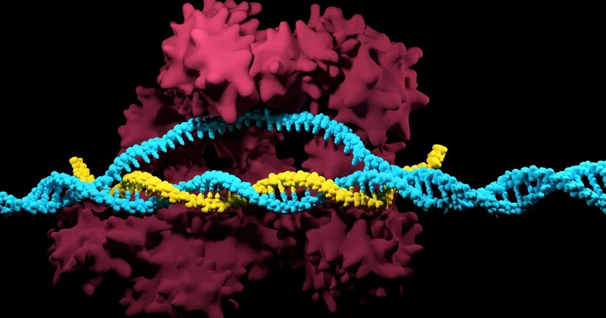
CRISPR/Cas9, short for “clustered regularly interspaced short palindromic repeats and CRISPR-associated protein 9”, was naturally developed by bacteria as a defense mechanism against invading viruses. As a part of this mechanism, bacteria confiscate fragments of DNA from invading viruses, and use the fragments to construct CRISPR arrays [1]. CRISPR arrays give the bacteria the ability to recognize identical or similar viruses, so if a similar virus attacks again, the bacteria produces RNA segments from the CRISPR array to specifically attack the virus’s DNA [1]. Cas9, an enzyme, is then used to cut apart the DNA, essentially killing the virus [1]. Scientists in the medical community are now adapting this gene-manipulation system to be used in cells in other organisms, including humans. The genome-editing tool has unlocked the potential to combat human genetic diseases previously deemed untreatable. A more cost-efficient, advanced, and precise method of gene-editing, CRISPR/Cas9 is revolutionizing the face of medicine as we know it [1].

Fascinated by the properties of CRISPR/Cas9 and microbes, Dr. Xue Gao, a professor of Chemical and Biomolecular Engineering at Rice University, currently investigates the applications of genome-editing and microbe-based natural product discovery. Her research “lies at the interface of chemical biology and biomolecular engineering with a primary focus on small- and macro-molecule discovery and their applications to human health, agriculture, and energy.” Dr. Gao’s contribution to the development of Statins, a pharmaceutical drug with lipid-lowering capabilities for those at risk of cardiovascular disease, piqued her interest in protein engineering. The frequent use of Statins in the medical society motivated Dr. Gao to continue synthesizing molecules to combat diseases. 

Her research also encompasses the use of enzyme-catalyzation (increase in the rate of a process due to an enzyme) for anticancer and antibiotic drug synthesis. Enzymes are powerful biological catalysts, accelerating chemical reactions by assisting in the formation of a transition state between the starting reagent and end product. Enzymes are also highly specific in which reactions it catalyzes, usually catalyzing only a single chemical reaction (or reactions very similar in nature), limiting side reactions and the ultimate formation of wasteful by-products [3]. According to Dr. Gao, whereas traditional chemical methods of synthesizing compounds require environmentally toxic solvents and harsh conditions, enzymes work well in moderate environments and are capable of producing a higher yield of 99%. As enzymes are proteins, extreme temperatures and pH levels result in denaturation, rendering the enzyme inactive. Therefore, enzyme catalyzation ability is enhanced at moderate conditions. In biological systems, a majority of processes utilize enzymes, from the transfer of CO2 in the body through the enzyme carbonic anhydrase, to breaking down sugars through the enzyme amylase. The development of enzymes to catalyze complex chemical reactions, called directed evolution, is a major focus of the Gao Laboratory. 

Dr. Gao ascribes “her love [for] exploring what has already been designed by nature” to the sheer intelligence of microbes and their methods of defense against viruses. “Microbes are super smart. They create protein encodings themselves to fight different types of environments.”  CRISPR/Cas9, another prominent example utilizing protein encodings and enzyme catalysis, has also attracted Dr. Gao’s interest. CRISPRs are specialized protein encodings, while Cas9 is an enzyme capable of cutting apart genetic material. Recently, Dr. Gao utilized the genome-editing capabilities of CRISPR/Cas9 to treat autosomal-dominant hearing loss. Autosomal-dominant diseases are inherited from an abnormal gene from one parent, even when the corresponding gene from the other parent is normal. Dr. Gao’s findings suggested that a targeted delivery of gene-disrupting agents such as CRISPR/Cas9 in vivo (within a living organism) can potentially treat such cases of hearing loss [2].

Dr. Gao’s research group is planning to do further work in CRISPR/Cas9 applications for a variety of human genetic diseases, and hopes to make breakthroughs in research through accommodating advanced computational tools and developing DNA sequencing technology (technology that determines the sequence of nucleotide bases). Dr. Gao envisions the next stage of her group’s research to be developing new enzyme-catalyzed reactions in the energy sector, particularly the production of biofuels, and combating food contamination in the agricultural industry. 

Dr. Gao’s current and future research aspirations in the field of human genetics and the energy sector serve as an inspiration for aspiring female scientists globally. She hopes that young females will follow their passion in STEM, and envisions a future where female scientists are not in the minority. Because of Dr. Gao and her vision, the Gao Laboratory is not only an embodiment of cutting-edge innovation in the future of enzyme-catalyzed reactions, but also the inspirational potential of female scientists. 

Works Cited

[1] What are genome editing and CRISPR-Cas9? - Genetics Home Reference - NIH. https://ghr.nlm.nih.gov/primer/genomicresearch/genomeediting (accessed Nov 8, 2019).

[2] Gao, X., Tao, Y., Lamas, V. et al. Nature. [Online] 2018, 553, 217-221. https://www.nature.com/articles/nature25164 (accessed Nov 8, 2019).
[3] Berg, J.; Tymoczco, John; etc. Biochemistry, 5th; W.H Freeman & Co: New York, 2002; 303-341.

Comment

Comment

The Solution Right In Front Of (or is it Inside?) Our Eyes

by Nithya Ramcharan

Antibiotics have for long been a prominent facet of medicine and are ubiquitous in households today due to their accessibility. At the earliest sign of a bacterial infection, we consume antibiotics with the confidence that it will be eradicated within a couple of weeks. However, this confidence has led to antibiotic abuse, a major issue in the medical field currently. For one, overusing antibiotics depletes the natural fauna populating certain organs like our intestines, making them more vulnerable to virulent bacterial strains. Naturally-occurring bacteria in our organs are a component of the immune system’s first line of defense and play a crucial role in immune system homeostasis and development [1]. Most importantly, antibiotic abuse has led to what some sources dub “the Antibiotic Resistance Crisis.” Antibiotics are often overprescribed when many of the infections’ pathogens are left unidentified, so out of caution, infections are assumed to be bacterial. 80% of antibiotics sold in the US are funneled into livestock for growth and prophylaxis [2].  As a result, bacteria are rapidly developing resistance to many commercial antibiotics and are returning more potent than ever. Bacterial genes can be transferred both to relatives and non-relatives through plasmids, circular DNA molecules in bacteria that can replicate independently of their host bacterial cell. This can confer resistance rapidly not only within a single population but also between different bacterial species [3]. Researchers are thus looking for alternatives to antibiotics to curb bacterial resistance and its catastrophic consequences.

One solution researchers have identified is a group of proteins occurring naturally within our bodies. Known as “antimicrobial peptides” (AMPs), these proteins are small, positively charged molecules that function within organisms as a natural immune response against microbes. Most commonly, AMPs can kill a bacterial cell through lysis; interactions between the positively charged clusters of an AMP and the negative charges in the phospholipid bilayer of a bacterium’s cell membrane form pores in the membrane, which ruptures the bacterium [4]. AMPs can also inhibit bacterial growth by targeting intracellular operations after permeating the membrane. They are present in organs in the human body most susceptible to infection such as the skin and the eyes [4]. Crucially, unlike antibiotics, bacteria have not developed solid resistance to AMPs, which is a key reason why AMPs offer a more attractive and effective method of treating bacterial infections. However, there is still little established research on how exactly AMPs neutralize bacteria. 

Professor Anatoly Kolomeisky of Rice University’s Department of Chemistry has been investigating the mechanisms of AMPs. His work started with the observation that antibiotic abuse can be attributed to people wanting to completely eliminate bacteria from their systems. His team developed a theoretical framework based on preexisting quantitative data to analyze how AMPs kill bacteria by considering bacterial growth as well as AMP entrance and inhibition rates into the bacteria as stochastic (randomly determined) processes. The model, in essence, consisted of a set of chemical reactions that would eventually lead to elimination of bacteria from the system. “We looked at the system as a sequence of states,” said Dr. Kolomeisky. “These states differed from each other only in the amount of AMPs.” In the occasion that the AMPs kill the bacterial cell, the corresponding state is removed. He called the model a “one-dimensional picture in a chemical space,” in the sense that it provided more of a black-or-white outlook: with certain factors, all the bacteria would either be killed or none of them would be killed. Although the model is simplified and every biophysical process is not accounted for, the model offers a convenient method for calculating the dynamics of the reaction, such as bacterial growth rate and bacterial clearance rate and the parameters that can modify these rates. Additionally, unlike other theoretical research on AMPs, this study takes into account the stochasticity of the processes of AMPs entering and inhibiting the cell [5]. This offers an effective means to further understand the mechanisms of AMPs that can be analyzed statistically and later compared with empirical data. 

Using the model, the team found out that the probability of inhibition and bacteria clearance rate have a positive relationship. Additionally, both processes of AMP entrance and inhibition of bacteria are equally important for AMPs to suppress bacterial growth. One finding is that higher heterogeneity in the AMPs facilitate faster entrance rates, while faster inhibition rates lead to lower heterogeneity among the types of AMPs. Controlling the heterogeneity in AMPs is key to balancing entrance and inhibition rates and thus achieving an optimal bacterial clearance rate [5]. Even though the stochastic model is simplified to fit the available information, the mean inhibition times predicted by the model are consistent with those observed in other studies [5]. 

Once these properties are understood, the applications of AMPs are numerous. They expand beyond inhibiting just bacteria and could suppress other pathogens. They can potentially be used in regulating cell division, facilitating wound healing, assisting in surgical healing, and suppressing cancer cells. AMPs activated by environmental factors such as pH have been developed for drug therapy. LL-37, an AMP found in humans, is one example of an AMP currently in commercial use. It has been identified to have bactericidal, anti-cancer, and anti-inflammatory properties [7]. Aside from medicine, they can be used in food production as natural food preservatives. One of them, pedocin PA-1 (produced by a diplococcus, a type of round-shaped bacterium), is used as a food preservative to prevent meat deterioration [6]. AMPs have also been identified as potential animal feed components to boost health and immunity in animal farming and aquaculture. AMPs with antifungal properties could be used as pesticides in the agriculture industry. 

However, much is left to discover about AMPs, as they are a diverse group of peptides that work differently depending on their environment; for example, some operate at the millimolar scale, while some require nanomolar concentrations to function. In addition, knowing how to optimize the entrance and killing rates is important in determining the ideal level of heterogeneity among the AMPs attacking the cell. Dr. Kolomeisky is advancing his current research on AMPs by investigating how using more than one type of AMP affects bacterial clearance. Implementing a “cocktail” mix, as he called it, of AMPs would kill bacteria more efficiently. Even though the rate of the reaction decreases as more types of AMPs are introduced, the probability of killing bacteria increases. He is also researching the dynamics of cancer development and cell cycle regulation, and could combine his research with AMPs to help better understand both processes. 

Once the mechanisms of AMPs are better known, they can revolutionize biomedicine. They can replace antibiotics in many medical treatments, thus mitigating the Antibiotic Resistance Crisis yet still allowing antibiotics to be used when needed. Reducing unnecessary usage of antibiotics will insulate them from the imminent threat of resistance developing in bacteria so rapidly. Beyond medicine, AMPs serve as better alternatives for inorganic chemicals used in food preservatives, growth promoters, and pesticides as they are naturally-occurring molecules that might be less likely to harm the organisms they aren’t programmed to target. In the nature of an archetypal journey of self-discovery, the solution to the antibiotic resistance crisis lay within us this whole time.


Comment

Comment

The Brains Behind Smart Wound Dressings

by Puneetha Goli


Did you know that nearly 1.35 million people die each year as a result of traffic crashes? Or that  injuries sustained from these accidents are the leading cause of death for people ages 5 to 29 (1)? Currently, those injured in accidents are required to wait for personnel to arrive and scope out the scene before any medical help from a wound care provider can be given. However, imagine if instead, in the time a person waits for emergency services, a “smart” band aid could be applied immediately to the wound, setting the wound until advanced medical follow-up is available. 

This is exactly what Dr. Swathi Balaji, an assistant professor of Surgery at Baylor College of Medicine, is working toward. Alongside Dr. Taiyun Chi, an assistant professor of electrical and computer engineering at Rice University, Dr. Balaji hopes to develop an artificial intelligence-assisted smart wound dressing to speed up the wound healing process. 

Dr. Balaji received a PhD in biomedical engineering from the University of Cincinnati after she fell in love with the applications of the field and recognized the impact she could make by combining her foundation in mechanical engineering with bioengineering.

“I came for a Masters in Mechanical Engineering at the University of Cincinnati,” said Dr. Balaji. “I loved the program, …[but] it became very obvious that we were [only] making small incremental changes.” Her Masters work served as a turning point in which she realized she wanted to enter a field where she could make a more long-term impact, leading her to pursue a PhD in bioengineering. “There was so much going on in terms of orthopedic research, biomaterials, regenerative tissue applications, so I was very fascinated with that,” said Balaji. 

The work that Dr. Balaji conducted as a PhD student on tissue engineering, as well as her subsequent work on understanding the regenerative mechanisms of fetuses has inspired her current projects at Baylor. The goals of her lab are to understand intracellular communications and how they translate into an individual’s response to injury, scarring, and wound healing. Through examining healing mechanisms in mouse models, Dr. Balaji has worked to establish a pipeline, ranging from basic investigations to more complex interactions with biomaterials to address her lab’s goals. 

Dr. Balaji’s interest in regenerative wound healing coupled with calls of action from the Defense Advanced Research Projects Agency (DARPA) to better understand what is occurring at the wound scene is what led to a collaboration between her and Dr. Chi and ultimately gave birth to the idea of this innovative smart wound dressing (2). 

Dr. Chi’s lab is working on the development of novel biology-microelectronics hybrid systems. The technology entails using advanced integrated circuits and microfabrication techniques - the same process of fabricating CPU, memory, and wireless transceivers chips in our cellphones and tablets (3) - to create miniature-sized electronic biosensors and bioactuators (4). Although the technology already exists, his sensors are unique in that they are one of the first that can record multiple biomarkers simultaneously and on a flexible membrane, ideal for human skin. Recognizing the potential interdisciplinary overlap, the two began discussing the possibility of integrating the research done in Dr.Balaji’s lab with Dr.Chi’s sensors in order to create a machine-learning system that would be capable of collecting information from a wound site and administering an individualized treatment. 

“It’s a very exciting project...,” said Dr.Balaji. “We started off with simple machine learning but the goal is to ultimately go into more advanced predictive modeling and make it smarter as we go on, and the dream would be ... to deploy this in the battlefield or when there is an emergency or when there is an accident - to be able to slap it on and let the wound healing happen right from the get-go.”

Currently, the project is in its initial stages. The teams have worked to perfect their respective independent technologies—Dr.Balaji’s team has developed their understanding of the science using animal models, while Dr.Chi’s team has refined the sensors and actuators. Now, they are currently working to integrate both of these components and apply them to a wound site. 

However, despite the preliminary nature of her work, Dr.Balaji already sees multiple avenues for the future of her research. One of her goals is to promote this niche field of science in an academic environment:  “I think our goal is to develop this new field of biology - machine learning microelectronics hybrid systems - either as a new course for undergraduate or graduate education or a new pipeline for promoting STEM-based education,” said Balaji. In addition to the educational angle, Dr. Balaji expressed an interest in getting the SMART wound dressing patented. In the future, she hopes for a world in which her SMART wound dressing could have a wide range of applications - from healing a scratch from a playground fall to something more complicated such as a burn wound. 

The potential of Dr.Balaji’s and Dr.Chi’s project has been recognized by the John S. Dunn Foundation Collaborative Research Award Program and the duo was awarded the 2020 John S. Dunn Collaborative Research Awards which was established to “fund projects with high potential for impacting human health”(5). “We were very excited..[and] we see a lot of potential,” Dr.Balaji said of winning the award. She believes the award will give her and Dr. Chi’s team the necessary resources to dedicate the time and effort to produce a final product that can be tested and examined. “I think at the current stage, we have all the different pieces of the puzzle, and over the course of the next two years, ...the Dunn Award is really going to help us bring our technology together. To come up with an idea that is received very well makes it a very, very rewarding experience.” 

References 

  1. Road traffic injuries https://www.who.int/news-room/fact-sheets/detail/road-traffic-injuries (accessed Feb 24, 2021). 

  2. Intelligent Healing for Complex Wounds https://www.darpa.mil/news-events/2019-02-06a (accessed Mar 3, 2021).

  3. https://www.lifewire.com/what-is-cmos-2625826

  4. https://www.nature.com/subjects/biosensors

  5. Funding Opportunities – Gulf Coast Consortia https://www.gulfcoastconsortia.org/home/research/funding-opportunities/ (accessed Feb 24, 2021).

Comment

Comment

X Marks the Spot: High-Specificity Alternatives to Small Molecule Drugs

by Jessica Cao

“Side effects may include headache, dizziness, nausea, and vomiting, and serious, sometimes fatal allergic reactions may occur.” Most common and modern-day pharmaceuticals, such as those seen in medical advertisements, are classified as small molecule drugs (SMDs). These are defined as drugs with low molecular weight and are “capable of modulating biochemical processes to diagnose, treat, or prevent diseases”. [1] The nature of SMDs makes them particularly attractive in the pharmaceutical industry, as they can be easily manufactured and regulated compared to more complex compounds. This ease and convenience in turn lowers costs, allowing them to be more accessible and affordable for patients. [2] Unfortunately, the simple composition of SMDs typically results in a litany of unwanted side effects due to their inability to target specific protein receptors.The physiological dangers of these side effects have spurred the scientific community to discover other, potentially safer methods of drug delivery. 

Among these researchers is Dr. Jerzy Szablowski, an Assistant Professor of Bioengineering at Rice University. Dr. Szablowski’s research focuses on neurotherapeutics, particularly the development of technologies that can noninvasively monitor and interact with the brain.3 One of the projects at the center of his research is gene therapy. Dr. Szablowski believes that this is a distinctly promising method of drug delivery “because it can be separated into different layers of specificity.” He states that, unlike small molecule drugs, where “everything is encoded within several atoms of the molecule”, gene therapy can be optimized for specificity and modularity. Gene therapy involves the delivery of a new gene to a cell through a viral vector and a promoter, which is a DNA sequence that helps facilitate the production of the protein encoded by the gene. This type of multifaceted system is advantageous because each component can be modified for different uses. Since the purpose of the viral vector is to deliver the gene (the therapeutic component), the vector can be modified to treat specific parts of the body without altering the treatment. Furthermore, Dr. Szablowski elaborates that “the promoter [of the gene] determines subtype, [which can] provide specificity.” Ultimately, the ability to modify each  component of gene therapy without affecting the others yields greater specificity for each drug. Greater specificity allows for the avoidance of side effects resulting from non-specific binding between the drug and body receptors that are unrelated to the disease being addressed. However, assessing neurological illnesses to determine necessary treatment is often difficult, as brain biopsies are highly invasive and risky. 

In order to gain a better understanding of the pathology of neurological disorders and the effects that they have on specific parts of the brain, Dr. Szablowski and his lab have employed a method of controlling and monitoring brain tissue via ultrasound. This noninvasive technique involves focusing ultrasounds on a specific part of the skull to open the blood-brain barrier (BBB) by a few millimeters. As Dr. Szablowski describes, the ultrasounds are delivered in conjunction with “a special contrast agent that is FDA-approved, [which is] basically a bubble that is encased in a lipid. Because this contrast agent has gas, it can actually become compressed or [expanded].” He further specifies that ultrasounds are essentially “an expansion and compression of a mechanical wave over time”, which pushes apart blood vessels in the brain, causing the [impermeable] junctions of the BBB to separate, and the endothelium (a thin membrane) in the brain to become more permeable. During this stage, a patient’s blood can be drawn, and a simple blood test can be used to assess their neurological condition.4 This procedure can alternatively be used to deliver therapeutic nanoparticles and proteins such as antibodies to specific parts of the brain. Although the concept of opening the BBB may sound risky, Dr. Szablowski assures that “several hours post-application, the impermeable junctions basically close up, and they become normal again.” Furthermore, little to no side effects have been observed apart from minor brain inflammation, which is easily treated with the steroid dexamethasone. Dr. Szablowski believes that this method of focused ultrasound can be used independently or in conjunction with gene therapy to ultimately become a common method of highly specific drug delivery to the brain. 

Dr. Szablowski also hopes to expand on his current projects to discover novel methods of low-cost drug delivery, such as non-genetic therapy. He mentions that non-genetic therapy involves the “delivery of proteins that actually produce the drug on site, and the production of the drug on site allows it to be specific.” In other words, the biological site(s) involved with a disease are the only sites being treated, with other parts of the body remaining unaffected. Furthermore, the Szablowski lab has recently begun the Therapeutic Improvement Project, which will potentially simplify the arduous and expensive process of drug development. Dr. Szablowki says that currently, when developing a drug, “you first have to make a cell model of the drug, [then] find what receptors are responsible for the disease, then test [using a] tissue culture and mouse or larger animal, and if you're lucky and pass through all those stages, you can [use on] humans.” Unfortunately, because animals and humans are biologically different, a drug can affect each organism differently, and projects often remain “stuck” at the animal model phase. Dr. Szablowski and his lab are hoping to bypass the many obstacles of drug development by creating “a single drug [that] can treat multiple diseases by just delivering to single cells.” 

Ultimately, Dr. Szablowski hopes that his research will be applied to a number of diseases—most notably neurological ones such as Parkinson’s, epilepsy, and PTSD—as opposed to a specific illness. Many of these diseases are currently difficult to assess, monitor, and treat in real time, but Dr. Szablowski believes that these minimally invasive, yet highly specific methods will eventually allow the development of therapeutic treatment with little to no side effects. 


[1] Ngo, H. X.; Garneau-Tsodikova, S. What are the drugs of the future? https://www.ncbi.nlm.nih.gov/pmc/articles/PMC6072476/ (accessed Feb 24, 2021). 

[2] Gurevich, E. V.; Gurevich, V. V. Therapeutic potential of small molecules and engineered proteins. https://www.ncbi.nlm.nih.gov/pmc/articles/PMC4513659/ (accessed Feb 24, 2021). 

[3] Szablowski, J. Jerzy Szablowski: The People of Rice: Rice University. https://profiles.rice.edu/faculty/jerzy-szablowski (accessed Feb 24, 2021). 

[4] Miller, B. Targeting ultrasound for noninvasive diagnosis of brain cancer: The Source: Washington University in St. Louis. https://source.wustl.edu/2020/08/targeting-ultrasound-for-noninvasive-diagnosis-of-brain-cancer/ (accessed Feb 24, 2021).


Comment

Comment

The Mighty Might-ochondria

by Pavithr Goli

Famously known as the “Powerhouse of the Cell”, the mitochondria is vital as it provides the human body with the necessary energy to function on a day-to-day basis. In addition to this main role, researchers have recently discovered other mitochondrial functions critical to human survival such as interacting with pathogens during infections and metabolizing cholesterol (1). These often overlooked features of mitochondrial function have intrigued Dr. Natasha Kirienko, an Associate Professor in the Department of Biosciences at Rice, who has been studying mitochondrial surveillance pathways and damaged mitochondria. Furthermore, Dr. Kirienko has spent time studying how both can be utilized in anticancer therapeutics, which can potentially change the approach taken to treat cancer, saving numerous lives in the process.

Dr. Kirienko joined Rice University in 2015 after she earned a $2 million grant from CPRIT (Cancer Prevention & Research Institute of Texas). After earning her undergraduate and graduate degrees in biochemistry and biology in Russia, Dr. Kirienko came to the United States to work on her Ph.D. After receiving her Ph.D. in molecular biology from the University of Wyoming, Dr. Kirienko worked as a postdoctoral research fellow at the Massachusetts General Hospital and Harvard Medical School before joining Rice to continue her groundbreaking research on mitochondria. Dr. Kirienko developed her fascination for mitochondria during her time in graduate school where she was interested in why “mitochondria had lower efficiency in leukemia” and why “they had a poor ability to convert electrons into ATP.” 

As someone who has “always [been] interested in identifying fundamental principles of why things are working,” Dr. Kirienko was curious about the effect of disease on mitochondria and, “the biochemical basis of the cancer cells /cell lines.” This interest in studying the pathogenesis of mitochondria propelled her to begin conducting research and investigating the response of damaged mitochondria and the strategies these damaged structures employ to recover when pathogens impact their function. 

Dr. Kirienko spent years dedicated to developing a greater understanding of mitochondria with the hope that a new therapeutic method to fight cancer could be developed. This interest in understanding fundamental principles led Dr. Kirienko to focus her research on one of the most fundamental and basic organisms that exist: Caenorhabditis elegans or C. elegans (2). A roundworm commonly used in scientific research as a model organism, C. elegans has been instrumental in understanding the various molecular and genetic mechanisms the human body employs when responding to diseases like cancer (2). 

Using this model organism and previous literature as a guide, Dr. Kirienko has been able to observe novel pathways in mitochondria and find drugs that induce mitochondrial damage, pushing “cancer cells [to] undergo cell death like paraptosis or apoptosis.” The goal of Dr. Kirienko’s current approach is to apply this novel method of inducing cell death on ''different cancer cell types” or combine it with current anticancer drugs in a synergistic manner to create highly selective treatments capable of killing cancer cells and sparing healthy ones. This approach will help reduce the side effects commonly seen in cancer treatment while maintaining the treatment’s effectiveness.

To further her research, Dr. Kirienko uses an interdisciplinary approach by working with physical chemists and doctors to help ensure that her novel cancer therapy will be applicable beyond C. elegans. She hopes that physical chemists can optimize the drugs that Dr. Kirienko found by “synthesizing and improving the physical structure of the drug.” After further testing on mice and cell cultures, Dr. Kirienko intends to expand her research and consult the help of physicians in a potential clinical study.

Along with her cancer research, Dr. Kirienko has expanded her lab’s focus on other diseases caused by poor mitochondrial health such as Alzheimer’s disease. Specifically, she and her lab are trying to understand whether improving mitochondrial health may be beneficial for neuronal function. In order to understand this mechanism further Dr. Kirienko is attempting to use various biomolecules to avoid cellular death by activating mitochondria and “[finding] a way to help mitochondria fix themselves quicker.” 

Furthermore, Dr. Kirienko and her lab are attempting to observe mitochondrial reactions to infections. Using C. elegans as their model organisms, they are interested in “looking at basic toxins that damage mitochondria.” One specific group of toxins that they are investigating are siderophores, which are “small molecules with a peptide side chain… [that] are secreted in the body and release iron.” During a bacterial infection, the bacterial pathogen uses its siderophore to absorb iron from the host, enabling the bacteria to grow and infect the cells. The mitochondria are the site of electron transport chains (ETCs), which contain several iron-rich proteins that help generate energy for the cell; unfortunately, when “iron is stolen, the mitochondria get damaged.” (3).To better study this mysterious process, Dr. Kirienko and her lab have “developed a cell culture model where [they] can assess the pathogenicity of bacteria.” They aim to use this newly developed model to elucidate the reactions and mechanisms that allow pathogenic bacteria to hijack cellular mechanisms.

Beyond discovering mitochondrial pathways and reactions to infections, Dr. Kirienko's lab is eager to explore "mitophagy or mitochondrial turnover, applied to cancer… [and] study more mitochondrial pathways.” She hypothesizes that this research can provide key information in finding ways to strengthen mitochondrial health so that mitochondrial cells can effectively withstand deadly pathogens and diseases, ultimately leading to longevity in human health. 

Dr. Kirienko and her lab have been working on some groundbreaking research for the past five years. Using novel methods and ingenuity, Dr. Kirienko has already made major strides in the field of cancer therapy and molecular biology. Through her current projects and the future endeavors that she hopes her lab can make, Dr. Kirienko intends to understand more about the powerhouse of the cell and use it to develop influential cancer therapies. 

Works Cited:

(1)Wallace, Douglas C. “Mitochondria and cancer.” Nature reviews. Cancer vol. 12,10 (2012): 685-98. doi:10.1038/nrc3365  

(2)Why Study C. Elegans?, Dr. Lundquist, 01/19/21, www.people.ku.edu/~erikl/Lundquist_Lab/Why_study_C._elegans.html

(3) Montine TJ, Morrow JD. Fatty acid oxidation in the pathogenesis of Alzheimer's disease. Am J Pathol. 2005 May;166(5):1283-9. doi: 10.1016/S0002-9440(10)62347-4. PMID: 15855630; PMCID: PMC1606384.


Comment

Comment

Evaluating the Implications of COVID-19 on Patients with Sickle Cell Disease

by Harveen Kaur

Abstract

The presence and rapid spread of the novel infectious disease COVID-19, caused by the respiratory pathogen/coronavirus SARS-CoV-2, has encompassed the world as a global pandemic. COVID-19 spreads contagiously through air and droplets of bodily fluids, such as saliva, and has an array of symptoms associated with its contraction and spread, leading to approximately 364 cumulative coronavirus-related hospitalizations per 100,000 individuals only in the United States during early 2021. Additionally, COVID-19 cases are often more severe in individuals that have pre-existing medical conditions, with sickle cell disease being at the top of the list. Sickle cell disease, also referred to as sickle cell anemia, is an inherited blood disorder that abnormally distorts the structure and function of hemoglobin in the body leading to oxygen deficiency. This medical condition has been associated with increasing the risk of developing a severe case of COVID-19, but a definite connection has not yet been confirmed that categorizes the risk factor for various age groups. Studies published in September 2020 report that an international registry, called SECURE-SCD, has been created to track patients suffering from both conditions and their respective health outcomes. By using the data from various documented studies, this article hypothesizes that there is an important connection between sickle cell anemia and COVID-19 that leads to enhanced symptoms and overall higher fatality in patients suffering from the two. Hence, the primary purpose is to dissect and evaluate the specific linkage between the SARS-CoV-2 coronavirus and sickle cell anemia including the risk factors for both medical conditions experienced simultaneously and based on key demographic categories. Establishing the connection between sickle cell disease and COVID-19 is significant to better understand the implications of the virus for those with pre-existing conditions and potentially adjust treatment protocols that are currently being discovered.

Introduction

Infectious diseases have proven to be the ‘silent killer’ of the world throughout history. Ranging from the Black Death bubonic plague, the Spanish flu, and the swine-origin influenza A (H1N1) virus, the slow but steady upward  trend of infectious disease occurrence  enforces the fact that a human-dominated world can never thrive with imbalances in nature [1]. Many of these infectious diseases are zoonoses, diseases in animals that are transmitted to human beings, while others are classified as viruses transmitted by human beings to other human beings [2]. Virulent microorganisms have been present  among humankind for thousands of years, but it is only within the past couple of centuries that they have caused a huge leap in mortality. The most recent example of a globally destructive pathogen is the SARS-CoV-2 coronavirus, which causes the respiratory syndrome [2]. 

COVID-19, a respiratory illness that the world has been facing as a pandemic for over one year, is a contagious infectious disease stemming from the SARS-CoV-2 respiratory pathogen with the most common symptoms as fatigue, cough, and fever [3]. According to the CDC, the virus primarily spreads through respiratory droplets that arise through coughing and/or sneezing [3]. However, recent studies and speculation have stated that transmission may be airborne due to the presence of SARS-CoV-2 viral RNA in air samples within droplet nuclei that stay infectious if left in the air over long distances and large amounts of time [4]. Currently, there is no definitive cure for COVID-19, but many countries including the United States have implemented methodologies to track and contain the virus, especially through vaccine development. Specifically, roughly 78 vaccines for COVID-19 are undergoing clinical trials, with seven vaccines (such as those developed by Pfizer-BioNTech and Moderna) already distributed for full use [5]. These highly infected countries have also encouraged individuals to socially distance themselves from others by at least six feet and wear masks, both of which help control daily cases and fatalities from COVID-19 [6]. Despite these preventative measures, the implications of COVID-19 have affected millions of individuals in the United States and on a global scale. Vulnerable populations that already suffer from pre-existing medical conditions are at high risk to develop and contract a severe case of COVID-19. One such diagnosis that has caused an uproar in the United States is sickle cell disease.

Sickle cell disease (SCD), also called sickle cell anemia, consists of a group of inherited disorders that changes the shape of red blood cells from disc-shaped to crescent-shaped when atypical hemoglobin is present [7]. Through this change, red blood cells are more prone to blocking blood flow and do not move easily in the body, which can lead to an onset of chronic conditions such as strokes and other pain crises [7]. With the spread of COVID-19, however, those suffering from SCD have been studied to have a higher rate of contraction, as well as more severe symptoms and outcomes when coupled with the virus [8]. For example, SCD patients with COVID-19 have a higher overall case fatality (10.9% fatality rate) than non-SCD individuals contracting the virus (3.3% fatality rate) [9]. Patients suffering from SCD have cardiopulmonary comorbidities that COVID-19 heightens, but the specific linkage between the two has yet to be closely compared. However, certain unmistakable trends connecting patient’s age, race and severity of SCD symptoms to their COVID-19 prognosis have been observed [8]. This article focuses primarily on the results from various national and international registries that collect information about COVID-19 and SCD by comparing the effects of suffering from both conditions together, based on age groups and other patient demographics. Establishing and characterizing the connection between the two conditions can create a more accurate solution on how to protect oneself from COVID-19 when suffering from SCD.  

Materials/Methods

The connection between SCD and COVID-19 transmission and symptoms, when specifically focusing on age and race, can be outlined through the data found in three studies. 

The first study, published on the Centers for Disease Control and Prevention, was conducted from March to May 2020 in the United States and was recorded in the Medical College of Wisconsin SECURE-SCD Registry [10]. This particular registry examines COVID-19 cases among those living with SCD in the United States, and the results of the study were limited to a two-month time period in which such patients were analyzed and documented. The study primarily focused on the average age of these patients, the number of SCD-related complications these patients have had in the past, and the resulting intensity of their current COVID-19 symptoms [10].

Another study, published in the journal Haematologica, referenced 10 patients in the UK who had contracted SARS-CoV-2, and all 10 patients had hemoglobin SS disease, which is the most common and most severe type of SCD [11]. The severity of this particular kind of SCD also means that these patients experience the worst symptoms at the highest rates for the condition [12]. With the most recent documentation being published in November 2020, the recovery rates of these patients were analyzed. The figure below from the same study highlighted the steps of evaluation conducted for the patients with SCD who had symptoms that aligned with potentially contracting COVID-19 as well as further standards of treatment and evaluation based on individual patient status [11]. The diagram shows the process of triage for patients with SCD who also contracted COVID-19 based on symptoms and patient accessibility [11].


Figure 1 Determining the appropriate medical response for SCD patients with COVID-19, ranging from patient instructions, types of medical evaluation, and potential therapies [11].

A third study, presented at the American Society of Hematology, was led by Dr. Lana Mucalo from the Medical College of Wisconsin and required the examination of 370 COVID-19 cases in patients already suffering from SCD from an international sickle cell registry [13]. This particular study compared the data found from the patients listed in the international registry to patients suffering from both conditions in the general Black population, keeping race as the measured demographic independent variable [13]. 

Results

The results of the first study, documented by the Medical College of Wisconsin SECURE-SCD Registry in between March 20, 2020 and May 21, 2020, showed that roughly 178 individuals who had SCD — and who were not simply carriers of the sickle cell trait — also contracted COVID-19 during this time. [10] Out of these 178 individuals, 7% (13 individuals) died upon contracting COVID-19 and being hospitalized, and 11% (19 individuals) were admitted to the intensive care unit while suffering from both conditions simultaneously [10]. More than half of the total individuals had suffered an SCD-related complication in the past that led to prior hospitalizations, and the average age of the 178 individuals at the time was roughly 28.6 years [10]. On the contrary, individuals with COVID-19 that don’t suffer from SCD have a death rate of less than 1% for those 20—54 years of age in early 2020, which is dramatically less than rates analyzed for patients with both SCD and COVID-19 [14].  

From the 10 patients in the UK documented in Haematologica, a 54-year-old patient died who suffered from delayed hemolytic transfusion reactions and also had high levels of CRP (C-reactive protein), a marker of negative prognostics for patients suffering from COVID-19 [11]. From all 10 patients, two underwent hydroxyurea therapy, seven had regular blood transfusions, and a total of five patients were managed remotely through telephone contact for their treatment [11]. Medical professionals and experts believed that suffering from the SARS-CoV-2 infection along with SCD would lead to acute chest syndrome (ACS). However, in the 10 patients, only one of them suffered from related respiratory complications; this was the same patient who passed away during clinical monitoring [11]. 

In the final study, the Black population was examined since, especially in the United States, individuals with African-American descent are statistically more likely to have both the sickle cell trait as well as the disease [13]. Additionally, completely separate from SCD, Black Americans are nearly four times more likely to be hospitalized due to COVID-19 as the virus has progressed globally [13]. Using race as the measured variable, this study concluded that those with SCD are roughly 6.2 times more likely to suffer a fatal complication from COVID-19 when compared to the general African-American population in the United States [13]. While the fatality rate based on ages 18-34 and 35-50 for the general Black population is less than 1% for all Black people in both age groups, those with SCD had death rates of 2.6% and approximately 12%, respectively, for the same age groups [13]. Additionally, a significantly larger amount of COVID-19 hospitalizations for Black individuals with SCD occurred among the younger age group, which correlates with larger death rates as age decreases [15].

Discussion

With the knowledge of all three pieces of data, these results illustrate that there is a correlation regarding age and race when examining the compounding effects of SCD and COVID-19. When examining the whole percentage of individuals who were hospitalized and severely affected by suffering from both conditions, the data from the SECURE-SED Registry suggests that such a connection exists. Additionally, data from the registry includes reports of 7% fatalities and 11% admittance to the ICU over 60 days for individuals with both conditions, which is heavily alarming. Even in the UK, the data suggests that there is an evident conjunction between the two conditions, despite the study being conducted with a small sample size. If 1 out of 10 individuals died, a 10% mortality rate from the combined detrimental effects of both conditions is just as concerning, if not more, than the first study. Lastly, the final study examined the effects of racial background on contracting COVID-19 while having SCD, and a significant relationship between the two is evident. In fact, having SCD, particularly for Black Americans, dramatically increases the fatality rate of these individuals from COVID-19 (by up to 11%). Based on these aforementioned studies, strong evidence exists to suggest that SCD and COVID-19 do have detrimental implications for individuals on a global level.

Conclusion

After understanding the symptoms of patients already suffering from SCD, adding the effects of COVID-19 can exponentially deteriorate a patient’s health. Through the three different pieces of data, there is a noticeable connection between incidence of SCD and severity of symptoms of COVID-19, the most recent and prevalent globally-spread virus. These connections can be analyzed on the basis of age as well as race, thereby indicating that the hypothesis for a linkage between the two conditions is well-supported. Since COVID-19 treatments and vaccinations are still being actively manufactured, knowing its added implications for those with SCD can be significant when determining a proper healing regimen for the virus. Additionally, understanding the damage done by both ailments combined can help reduce their high mortality rate, thereby creating more successful outcomes for patients for both diseases individually and together. 

Works Cited

[1] Bean, Mackenzie. Becker’s Hospital Review. 2020. 

[2] Tabish, Syed Amin. International Journal of Health Sciences. 2009, 3(2) V-VIII.

[3] Coronavirus Disease 2019 [Online]. 2021. https://www.cdc.gov/dotw/covid-19/index.html (accessed Mar. 17, 2021).

[4] Liu, Jiaye. Emerg Infectious Dis. 2020, 26(6), 1320-1323.

[5] Zimmer, Carl. The New York Times. 2021.

[6] Thu, Tran P. B. Elsevier Pub Health Emerg Cond. 2020, 742.

[7] National Heart, Lung and Blood Institute. US Dept. of Health & Human Services. 

[8] Shet, Arun. American Society of Hematology. 2021.

[9] Minniti, Caterina P. Blood Advances. 2021, 5(1), 207-215.

[10] Panepinto, Julie A. Centers for Disease Control and Prevention. 2020.

[11] Menapace, Laurel A. Haematologica. 2020, 105 (11), 2501-2504.

[12] Rogers, Graham. Healthline. 2019. 

[13] Thompson, Dennis. Medical Xpress. 2020

[14] Razzaghi, Hilda. Centers for Disease Control and Prevention. 2020, 69(12), 343-346.

[15] Mucalo, Lana. American Society of Hematology. 2020.

Comment

Comment

Determining the Efficacy of Sourcing IgY Antibodies from Chicken Eggs Using the PierceTM Chicken IgY Purification Kit

by Sachi Kishinchandani

ABSTRACT

IgY is known to be an immunoglobulin that is formed by the maternal immune system and passed down to offspring, in reaction to certain foreign substances. It is hypothesized that IgY can be used to boost human immune systems. Thus, to ask how we can easily gather these proteins for increased human immunity, we analyzed the purification of IgY via the Thermo ScientificTM PierceTM Chicken IgY Purification Kit. Through a Bradford assay, SDS PAGE, and a Densitometry, we determined that it is possible to purify around 40-80 mg of IgY per egg using similar methods. Since each hen produces hundreds of eggs annually, and each egg contains such a large portion of IgY, it is reasonable to conclude that mass purification of IgY is feasible.

INTRODUCTION/BACKGROUND

Our immune system plays a vital role in protecting our body from foreign diseases, viruses, and harmful bacteria. Therefore,the study of its mechanisms and functions is extremely important. One important feature of the immune system across a variety of species is how antibodies are passed from mothers to their newborn offspring [1]. Over time, natural selection has led to adaptation of antibodies to be passed down to offspring for survival, whether it is in the form of the IgG antibodies passing through the placenta in mammals, or birds passing down IgY antibodies in their egg yolk [1].

By studying the IgY antibodies found in chicken eggs, we can study and potentially introduce useful IgY antibodies into humans (for example, via the consuption of IgY Antibodies), in order to bolster our immune systems. IgY antibodies can be used to create immunization in a form that does not specifically require a vaccine [2]. People can be passively immunized against respiratory illness with the help of IgYs to help neutralize respiratory pathogens and disease spread. IgY is an ideal candidate for this antibody insertion due to its structural similarity to the IgG antibodies common in humans, as well as its ability to target specific antigens [3]. IgY has been found to bind specifically to influenza virus Nucleoprotein (NP) by detecting multiple virus subtypes in swine [4]. These traits make IgY a worthwhile subject of study, especially one whose potential outcomes are so beneficial. 

In this series of experiments, we focused on investigating IgY protein concentrations, abundance, and purity in potential sources for IgY protein purification, as the extent of these properties are not widely available in current literature. We used the Thermo ScientificTM PierceTM Chicken IgY Purification Kit to extract the IgY protein from the egg yolk [5]. The extraction of the IgY protein allowed us to conduct a Bradford assay, SDS PAGE, and a Densitometry in order to determine the concentration, abundance and purity of the protein. With the results of this experiment, we determined that we can effectively purify IgY from store-bought chicken eggs.

MATERIALS AND METHODS

PHYSICAL PURIFICATION OF IgY PROTEIN

We purified IgY from store-purchased Eggland Best eggs using the Pierce™ Chicken IgY Purification Kit from Thermo Fisher to physically separate the antibody rich yolk from the white using a series of suspensions and centrifugations with reagents. We conducted this by first separating the egg yolk from the egg whites. We then rolled the egg yolk on a paper towel to remove excess egg-white protein, pricked the yolk sac and then drained an approximate 8 mL of the yolk into a beaker. This step is essential to remove any contaminant protein from the egg white. We then added five times the egg yolk volume of cold Delipidation Reagent (40 mL) from the Thermo ScientificTM PierceTM Chicken IgY Purification Kit to the 8 mL egg yolk. The mixture was stirred until well incorporated and then stored at 4 ºC for a week. 

The mixture was then centrifuged for 15 min at 6,000 × g in a refrigerated centrifuge. The colorless and translucent supernatant was decanted and placed in a secondary conical tube, to which the precipitation reagent solutions from the Thermo ScientificTM PierceTM Chicken IgY Purification Kit were added. The mixture was placed in a 4 ºC incubator overnight, and then centrifuged for 15 min at 6000 rpm in a 4 ºC refrigerated centrifuge. We then isolated the pellet from the conical tube.

Our final step in the purification of the protein was to resuspend the pellet in Phosphate buffered saline (PBS) (100 mM Na3PO4, 150 mM NaCl, pH 7.2). We added the PBS to the pellet and stirred the mixture until it was well incorporated. 


BRADFORD ASSAY

Our next step was determining the concentration of the IgY protein using a Bradford assay. The assay uses acidic Coomassie dye to bind to proteins and result in a visible color change from brown to blue, the intensity depending on the amount of protein present. We measured out 5 mL of Bradford reagent into 8 glass test tubes. We pipetted Bovine Serum Albumin (BSA) protein into the first six test tubes, and IgY into the remaining two test tubes. With the BSA protein test tubes, we measured the absorbance at 595 nm for each solution with a spectrophotometer, and created a standard curve of the absorbance in relation to BSA protein amounts. We then measured the absorbance of our protein to be 0.383. 


SDS PAGE

Because current literature information on IgY proteins states that two chains of IgY should occur around 70 kDa and 30 kDa [6], we decided to make our gel a 12% acrylamide. With our SDS PAGE, we first created a standard mixture of 1 L Running buffer, 10 mL of 1x Separating buffer and 10 mL 1x Stacking buffer. We also created a 1 mL of 10% APS solution for activating the polymerization of the gel by dilution of stock APS available in the lab. We then added 100 uL of APS and 10 uL of TEMED into both the Stacking and Separating buffers. We then began to pipette the Separating buffer solution in between the glass plates until a marked line, allowing the Separating buffer to set with isopropyl alcohol before adding the Stacking buffer on top. While the Stacking buffer was still liquid, we inserted the comb to create the wells in the gel. 

We stored the gel for four days in a moist paper towel, then conducted SDS PAGE with 5 uL of our protein and 5 uL of sample buffer in each of our wells, with the Broad Range 5 Microliters NEB in one of the wells for reference. We let the SDS PAGE run for 90 min --the first 10 min at 120 V and the rest of the time at 180 V--until the bands reached the bottom of the gel, and then stained the gel with Coomassie blue solution. We then destained the gel using the premade solutions Destain #1 (50% methanol, 10% acetic acid) and Destain #2 (7% methanol, 10% acetic acid). 

DENSITOMETRY

This part of the experiment was to determine the concentration of the IgY bands. The densitometry was conducted in a similar fashion to the SDS PAGE. In order to address the possible presence of a dimer in our previous SDS-PAGE gel, we increased the concentration of BME in two of the wells with our IgY protein in the hopes of removing any remaining disulfide bonds. Instead of creating a gel for this experiment, we used a BIO-TEC premade gel. We created 5 solutions of different concentrations of BSA: 0.2 ug, 0.5 ug, 1 ug, 1.5 ug, and 2 ug. We then added 5 uL of these solutions, mixed with 5 uL 2x Laemmli buffer into separate wells. We also added our purified IgY protein into the gel, with two wells made with 5 uL of protein and 5 uL 2x Laemmli buffer each, and two wells with 5 uL protein and 5 uL 10% BME Laemmli buffer each. We ran the gel at 300 V for 15 min, until the dye front hit the bottom of the gel. We stained and destained the gel exactly as we did for the SDS PAGE. We then used a machine to analyze the concentrations and Molecular Weight (MW) of the bands of IgY in comparison to BSA.

RESULTS & DISCUSSION

The overall purpose of the experiments was to determine the practicality of extracting large quantities of protein from store-bought chicken eggs. From the experiments, we were able to determine the following:

PHYSICAL IgY PURIFICATION

The weight of our purified protein match the expected result of using the kit. We were able to purify 70.2756 mg of IgY solution from a single store-bought egg (Table 1).

Table 1. The total mass of IgY protein solution as a result of the initial purification of IgY protein with Thermo ScientificTM PierceTM Chicken IgY Purification Kit. 

BRADFORD ASSAY

Once our protein was purified, we were able to determine the concentration of the protein from a Bradford assay, comparing the absorbance of our protein to the absorbance of BSA. 

Figure 1. The absorbance of BSA over the concentration of BSA measured via a spectrophotometer, used as a standard curve to determine that the concentration of our IgY protein is 2.43 ug/uL.

Table 2. Calculated concentrations of our IgY solution as determined by a Bradford Assay. Conducted by measuring the absorbance of different volumes of the IgY solution in a spectrophotometer. These data were used to find the average concentration of the IgY protein to be 2.43 ug/uL. 

With these calculated concentrations (Table 2), we found that the average concentration of our IgY sample is 2.43 ug/uL. This matched expected results as previous studies have estimated that the concentration of IgY purified from egg yolk should be 2-4 mg/mL [7]. 

SDS-PAGE

Knowing the concentration of the protein helped us conduct SDS-PAGE, which would help us find out the size and purity of our purified IgY protein. 

Figure 2. SDS-PAGE of our IgY protein with clear bands around 27 kDa and 130 kDa. Done in a 12% acrylamide gel that was stained with Coomassie blue solution. The left-most well on the right end of the figure indicates standard protein molecular weights from the NEB Protein standard solution. The remaining wells have 5uL of 2.34 ug/uL of IgY and 5uL of 2X Laemmli buffer, as indicated at the bottom of each column in the figure.  

Figure 3. Graph of the log (molecular weight) of NEB Protein Standard as a function of relative distance. The curve between the distances 2.5 and 4.4 cm was used to find the MW of the heavy band to be 117.7 kDa. The curve between the distances 14.7 and 18.6 cm were used to find the MW of the second band to be 27.28 kDa.

Once we conducted our SDS-PAGE and stained/destained it, we found that the gel showed significant IgY purity because the most significant bands were at around 27 kDa and 130 kDa. We also found a light band around 43 kDa (Figure 2), which was confirmed to be the contaminant ovalbumin by our instructors (see acknowledgments). 

We calculated the MW of the bands to be 27.28 kDa and 117.7 kDa. In order to do so, we calculated the equation of the line between the points 2.5 and 4.4 cm to be logMW = -0.7169 x + 2.2932. When we plug 3.1 in for x, the MW ends up being 117.7 kDa. We also calculated the equation of the line between points 14.7 and 18.6 cm to be logMW = -0.02987x + 1.971. Plugging in 17.9 for x, we get MW = 27.28 kDa (Figure 3). 

We expected to have the band at 27.28 kDa for the IgY light chain, but our other expected value for the IgY proteins was around 70 kDa for the heavy chain, not 117.7 kDa, based on current literature on IgY. Because this 117.7 kDa band was almost exactly twice the weight of our expected heavy chain weight, we hypothesized that our protein’s disulfide bonds had not broken down completely. In order to understand whether this high molecular weight was a result of the small amount of BME in the 2x Laemmli solution, or was simply a contaminant, we conducted a densitometry with two of the wells containing our protein along with a higher concentration of BME. 

DENSITOMETRY

Figure 4. The results of our Coomassie stained and de-stained IgY and BSA Densitometry [left-most well: NEB marker. Well 2-3: 10% BME + IgY. Well 4-5: IgY + normal Coomassie. Wells 6-10: decreasing concentrations of BSA] The red arrow points to the heavy IgY chain, which is a band we did not observe in the previous (SDS PAGE) experiment. The blue arrow points to the light IgY chain. The yellow arrow points to the BSA chain. 

SDS-PAGE was run, this time with more BME and varied BSA amounts to both analyze the light/heavy chains and to possibly get a reading of the bands that would help us determine the concentration of protein in each band. Figure 4 shows there is clearly a large amount of BSA in wells 6-10, which would have been difficult to analyze our IgY density with, so we were unable to carry out analysis of the IgY. 

We found that the gel with the higher percentage of BME had a more prominent band around 72 kDa, along with bands around 27 kDa and 130 kDa. The finding of a band around 72 kDa and 27 kDa was consistent with current literature on IgY protein chains. The bands around 27 kDa and 130 kDa were consistent with our previous SDS PAGE (Figure 4). We still assume that the band around 130 kDa could be the IgY protein’s heavy chain that remains a dimer, however, there is a possibility that this could be a contaminant.  

This experiment does not tell us the identity of each of the protein bands found in the densitometry. If we had more time, we would have run the experiment again, using lower BSA concentrations and using the BSA band densities to make a standard curve in order to determine the total integrated density of the protein. From there, we would have calculated the density and concentration of the IgY, which would have told us the purity of the protein in relation to BSA. However, since we were unable to conduct this experiment, we suggest that further experiments should be performed to determine whether the IgY was effectively purified. 

CONCLUSION

Based on preliminary results, we can reasonably conclude that our IgY purification was successful and we were able to extract around 24.3 mg of pure IgY protein from the 8 mL of store-bought chicken eggs. Current literature indicates that the expected concentration of protein from each egg yolk is 2-4 mg/mL, which was what we observed in our results. Other studies using similar methods to our experiment have also found that it is possible to purify around 40-80 mg of IgY per egg using similar methods [7]. The reason we predict 40-80 mg per egg yolk is because we only extracted 8 mL of yolk, whereas the average egg yolk is about 15 mL, [8] so the total protein amount would be around 40 mg for 15 mL of yolk. The 24.3 mg of IgY extracted from one egg shows that eggs are a reliable source of IgY protein. Since each hen produces hundreds of eggs annually, and each egg contains such a large portion of IgY, it is reasonable to conclude that mass purification of IgY is feasible. 

The extraction of IgY has important implications for further application. In the future, we hope to see multiple pathways for immunization built from IgY. One is the immunization of humans through digestion of IgY antibodies [9]. Another is epitope mapping, [10] identification and characterization of antibody binding sites on cells, which could help us understand the structures of antigen binding sites to combat diseases. The results of our purification of IgY seem promising for the future of IgY extraction and execution of further research. 

ACKNOWLEDGEMENTS 

We thank Dr. Thomas and Dr. Catanese for all their support and mentorship throughout the course. OURI, CUR, and the Biosciences Department for funding BIOS 211.  Anika Sonig and Aaron Lin for their partnership through this project and aid with the creation of the figures. BIOS 211 TAs for their guidance. 

Works Cited

(1) Pereira, E P V et al. International immunopharmacology 2019 vol. 73, 293-303. doi:10.1016/j.intimp.2019.05.015  

(2) Aymn Talat Abbas, Sherif Aly El-Kafrawy, Sayed Sartaj Sohrab & Esam Ibraheem Ahmed Azhar, Human Vaccines & Immunotherapeutics. 2019, 15:1, 264-275, doi: 10.1080/21645515.2018.1514224  

(3) Nagaraj et al. International Journal of Food Microbiology. 2016, 237, 136-141.

(4) da Silva M.C, Schaefer R, Gava D, Journal of Immunological Methods. 2018, 461, 100-105. https://doi.org/10.1016/j.jim.2018.06.023. 

(5) ThermoScientific PierceChicken IgY Purification Kit manual https://assets.thermofisher.com/TFS-Assets/LSG/manuals/MAN0011404_Pierce_Chicken_IgY_Purifi_UG.pdf

(6) Sudjarwo, S. A., Eraiko, K., Sudjarwo, G. W., & Koerniasari, Journal of advanced pharmaceutical technology & research. 2017, 8(3), 91-96. https://doi.org/10.4103/japtr.JAPTR_167_16 

(7) Pauly, Diana, et al. Journal of Visualized Experiments. 2011, www.ncbi.nlm.nih.gov/pmc/articles/PMC3197133/.    

(8) Egg Cooking Tips: Cooking With Eggs: Egg Farmers Of Alberta. eggs.ab.ca/eggs/egg-cooking-tips/. (Accessed November 24, 2021)

(9) Constantin, C.; Neagu, M.; Diana Supeanu, T.; Chiurciu, V.; A. Spandidos, D, Exp Ther Med, (2020), 20, 151–158. https://doi.org/10.3892/etm.2020.8704. 
(10) Lu, Y.; Wang, Y.; Zhang, Z.; Huang, J.; Yao, M.; Huang, G.; Ge, Y.; Zhang, P.; Huang, H.; Wang, Y.; Li, H.; Wang, W, Journal of Immunology Research (2020). https://doi.org/10.1155/2020/9465398.

Comment

Comment

Combatting E. Coli through Secretion Inhibitors

by Akshay Sethi

Introduction:

Escherichia coli (E. coli) is a rod-like anaerobic bacterium that infects the gastrointestinal tract of humans (Fig. 1).

Fig 1: (E. coli bacterial cells)

Most E. coli are harmless, but there are pathogenic strains such as Shiga toxin-producing E. Coli (STEC) that can cause diarrhea, abdominal cramping, and/or acute renal failure. [1] E. coli is found to colonize the human body hours after childbirth and does not pose harm unless the gastrointestinal barrier is broken or the host is immunocompromised. The microorganism is a threat to humans. Gram-negative bacteria possess cell envelopes and an outer membrane that can protect the inner membrane and its organelles from antimicrobial enzymes and antibiotics (Fig. 2). [2]

Fig 2: (Gram-negative bacteria have more components, including an outer membrane, to protect it)

The combination of a low concentration of E. coli needed to infect the host and its resistance to antibiotics makes the microorganism dangerous. [3]. E. coli has a core genome, or series of DNA strands that are common in all strains, but the bacteria also has a non-shared gene pool that differentiates each strain from anotherThere are at least six types of deadly pathotypes of E. coli., all of which cause enteric disease, or diseases relating to the gastrointestinal tract, and potentially lead to long-term injury. 

E. coli is not highly transmissible, as the risks come from contaminated food such as undercooked meat, unpasteurized milk, dirty water, or contaminated stool. [4] Therefore, the best prevention for the risk of infection is cleanliness such as washing one’s hands, as an individual must ingest the pathogen to contract it. [4] 

Novel therapries and treatments for E. coli have shown limited success. [8] Much of the successful experimentation has come from the process of injecting fluid with secretion inhibitors to stop production of key components of the bacterium. The main system for infecting host cells, T3SS, is the main target of secretion inhibitors and has been the focus of modern research on E. coli. [10]

Discussion:

Harms in E. coli:

 E. coli varies in its threat to humans based on the amount of Shiga toxins it produces. [3] Shiga toxins act as ribotoxins that inhibit protein synthesis, leading to altered gene expression or even apoptosis. This would lead to bloody diarrhea, or worse, the Hemolytic Uremic Syndrome (HUS). [5] HUS is one of the more dangerous outcomes of contracting harmful E. coli, where infected people suffer red blood infection and potential total kidney failure. [5] Global estimates project about 10% of STEC infections lead to HUS. HUS is the most common cause of acute renal failure in children, one of the two high-risk groups to contracting an E. coli infection. [5] The syndrome can also cause neurological failures in 25% of cases, resulting in symptoms such as seizure, coma, or stroke. [5]  

Shiga toxins are found in over 200 strains of E. coli. [7] Shiga toxins found in E. coli are found in the pathogenic serotypes of E. coli as Stx1. [4] A serotype is a specific group of bacteria sharing a common antigen, which is an immunogenic particle that incites an antibody response. Another serotype of E. coli contains  Stx2, which is a more potent Shiga toxin and a part of a different antigen serogroup. A serogroup consists of microorganisms differing in their composition of antigens. [4]  The Stx toxin is comprised of two key subunits—the A subunit and B subunit. The A subunit’s function is to inhibit protein synthesis by damaging the ribosome in an infected cell. [4] The B subunit binds to the globotriaosylceramide (Gb3), which is found in endothelial cells lining cardiac muscles and blood vessels. [4] Among biological substances, Shiga toxins are amongst the most poisonous and are lethal to various animals in small doses. E. coli carrying the Shiga toxin serotypes have been shown to lead to the worst outcomes of contracting the pathogen. Across different species, the Shiga toxins have been found to cause acute renal damage and in some cases, total renal failure. [7] Within population-based observations, along with experimentation on mice and baboons, Shiga toxins have been linked to HUS. [4] 

The pathway for Stx serotypes of E. coli to lead to HUS begins with the consumption of contaminated substances. E. coli rapidly replicates within the host and the Shiga toxins latch onto Gb3 before being enveloped and packaged into a microvesicle. This vesicle is then absorbed into the kidney endothelial cells, where the toxins cause necrosis, eventually leading to acute renal failure (Fig. 3). [6] 

Fig 3: (Stx binding to the GB3 on the cell membrane to take over the ribsomal components of the cell)

Besides the kidney, the brain is the second most affected organ in severe E. coli infection. The Shiga toxins attack neurons within the brain and can cause damage or partial failure to the central nervous system. Central nervous system damage can lead to seizure, shock, or paralysis in some cases. [7] 

Classifications of Shiga toxins go more in-depth than Stx1/Stx2 to distinguish between the potency of the toxin and the binding receptor. [7] Some types of Stx can cause severe loss of body mass, along with renal failure. [7] One common symptom of E. coli infection shared between most classifications is colon damage. Shiga toxins inhibit the function of the large intestine, which in turn causes severe symptoms such as bloody diarrhea. [7]

In the past couple of decades, Shiga toxin-carrying E. coli has been found in animal-to-animal transmission. [7] In an isolated study, STEC infection was found in zoonotic transmission, and a common source was domestic animals, such as house cats and dogs. Other sources included wild animals, but at similar rates. Most commonly found were feral hogs and hyenas. Scientists have looked to treat E. coli because of the long-lasting impact of the infection on the human body, including high blood pressure, heart disease, or kidney problems. [1]


Secretion Inhibitors:

One of the newly discovered methods in treating E. coli is anti-virulence therapy. This therapy takes advantage of the type III secretion system (T3SS) in E. coli that primarily exists to grow and multiply the bacteria within hosts. [8] The T3SS acts as a needle that will “inject” effector proteins into the epithelial cells of the gastrointestinal tract. Effector proteins are biomolecules that can attach to proteins and affect the production and type of product that the protein will create. These proteins function as ligands as well as secretory proteins, attaching mainly to host cells to infect them. The ligands will lower the activation energy to conduct the process of making bacterial cells, and therefore increase the rate of production. [8] The impetus for targeting T3SS by scientific researchers is that it is the main mechanism that both Enterohemorrhagic E. coli (EHEC) and Enteropathogenic E. coli (EPEC) use to infect and populate the host effectively. EHEC is the strain of E. coli that can cause HUS, while EPEC is less severe in bodily harm but is the leading strain for diarrheal deaths. [8] 

The method in which the treatment is proposed to work is unique. Instead of regulating or limiting the growth of new cells, the anti-virulent treatment targets and inhibits a virulence factor. [8] Virulence factors are what cause bacteria to cause disease in eukaryotic, multicellular organisms. [8] These factors will include molecules that will aid bacteria in infecting the host organism’s cells. [12] The factors are categorized as either secretory, membrane associated, or systolic. Systolic factos will lead to the bacteria changing its physiological or physical structure. [12] Secretory virulence factors assist the bacterium in counteracting the host’s immune response by releasing chemicals. [12] 

One inhibitor of the T3SS is salicylidene acyl hydrazide, which has been shown to be effective against EPEC, EHEC, and Salmonella. [9] Although effective, the salicylidene acyl hydrazide was discovered to indiscriminately bind to human proteins, which may negatively affect patient metabolism. [10] It is important to note that the effect of salicylidene acyl hydrazide on T3SS is broadly accurate in preventing the secretion system from functioning. [10] 

One important study explores the ways a secretion system inhibitor, Aurodox, can affect the T3SS function in different bacteria. [8] Aurodox is the novel system that incorporates salicylidene. [8] Although it does not affect the growth of bacteria, Aurodox has been shown to inhibit the secretion of T3SS. The effectiveness of Aurodox on inhibiting the T3SS system depends on the concentration of the Aurodox injected into the in vivo sample. [8] Researchers have found that in key strains of E. coli, Aurodox did not inhibit growth of the bacterium while inhibiting the mechanism of T3S production (Fig. 4). [8 ]

Fig 4: (Aurodox will inhibit T3S secretion, but not the growth of different bacterial strains)

In the study, researchers found that Aurodox has the function of inhibiting EHEC to infect epithelial cells in infected mice. Although results were inconclusive at the cellular level, Aurodox was shown to reduce colon damage in the mice. [8] The expression of T3SS in various proteins involved in DNA-binding and altering were also shown to have been inhibited by Aurodox. 

Conclusion:

These results are promising for the inhibition of T3SS expression in anti-virulent treatment. As noted previously, past antibacterial treatments had induced the SOS response in the EHEC strain of E. coli, which was not the case for the anti-virulent Aurodox. [8] This is significant because the SOS response as a function leads to the release of Shiga toxins. Without the release of the deadly toxins, EHEC infection can be inhibited.

Aurodox as a treatment poses an interesting future for combatting E. coli. While Aurodox can effectively inhibit the secretory function of E. coli through the T3SS system, it does not halt production of the bacterium. This poses a question of whether to accept the cohabitation of EHEC and EPEC strains, among others that are considered harmful in the present. [8] If Aurodox can effectively neutralize the harmful effects of the strains, then there is little reason to eradicate the bacterium from the host. 

Drawbacks:

One drawback to current antibiotic treatments which anti-virulent therapy attempts to solve is the SOS response by E. coli. [8] The anti-virulent therapy can cause minor DNA damage, signaling an SOS response. The SOS response will occur after the use of antibiotics and eventually lead to the overproduction of Stx in the intestines. This uptick in Stx production will lead to a higher chance of severe symptoms of the anti-virulence treatment. 

Antibiotic drawbacks are more feared by the scientific community because E. coli is the most common pathogen in humans. In a study analyzing 150 different food samples to test the antibiotic sensitivity pattern of the bacteria, the highest percentage of drug-resistant E. coli was found in the most common foods with E. coli present, which included raw meat, eggs, and salad. [11] To combat this, scientists propose that the general population practice good hygiene, and farmers should only use reserve antimicrobial drugs to reduce the possibility for antimicrobial resistance. [11] Anti-virulence treatment does not deal with antimicrobial drugs, and is more likely to reduce unwanted side effects of general treatment of E. coli as compared to other proposed solutions. 

[1] E. coli. (2018). Retrieved 20 December 2021, from https://www.who.int/news-room/fact-sheets/detail/e-coli

[2] Kaper, J., Nataro, J., & Mobley, H. (2004). Pathogenic Escherichia coli. Nature Reviews Microbiology, 2(2), 123-140. doi: 10.1038/nrmicro818

[3] Fact Sheet: Escherichia coli - Microbial Identification - MALDI ToF. (2021). Retrieved 20 December 2021, from https://wickhamlabs.co.uk/technical-resource-centre/fact-sheet-escherichia-coli/

[4] Melton-Celsa, A. (2014). Shiga Toxin (Stx) Classification, Structure, and Function. Microbiology Spectrum, 2(4). doi: 10.1128/microbiolspec.ehec-0024-2013

[5] Gram-negative Bacteria Infections in Healthcare Settings | HAI | CDC. (2021). Retrieved 20 December 2021, from https://www.cdc.gov/hai/organisms/gram-negative-bacteria.html

[6] E. coli: What is It, How Does it Cause Infection, Symptoms & Causes. (2021). Retrieved 20 December 2021, from https://my.clevelandclinic.org/health/diseases/16638-e-coli-infection

[7] Kim, J., Lee, M., & Kim, J. (2020). Recent Updates on Outbreaks of Shiga Toxin-Producing Escherichia coli and Its Potential Reservoirs. Frontiers In Cellular And Infection Microbiology, 10. doi: 10.3389/fcimb.2020.00273

[8] https://doi.org/10.1128/IAI.00595-18

[9] Zambelloni R, Marquez R, Roe AJ. 2015. Development of antivirulence compounds: a biochemical review. Chem Biol Drug Des 85:43–55.

[10] Dai Wang, Caroline E. Zetterström, Mads Gabrielsen, Katherine S.H. Beckham, Jai J. Tree, Sarah E. Macdonald, Olwyn Byron, Tim J. Mitchell, David L. Gally, Pawel Herzyk, Arvind Mahajan, Hanna Uvell, Richard Burchmore, Brian O. Smith, Mikael Elofsson, Andrew J. Roe, Identification of Bacterial Target Proteins for the Salicylidene Acylhydrazide Class of Virulence-blocking Compounds*, Journal of Biological Chemistry, Volume 286, Issue 34, 2011, Pages 29922-29931, ISSN 0021-9258, https://doi.org/10.1074/jbc.M111.233858. (https://www.sciencedirect.com/science/article/pii/S0021925819760700)

[11] Rasheed MU, Thajuddin N, Ahamed P, Teklemariam Z, Jamil K. Antimicrobial drug resistance in strains of Escherichia coli isolated from food sources. Rev Inst Med Trop Sao Paulo. 2014;56(4):341-346. doi:10.1590/s0036-46652014000400012

[12] Sharma AK, Dhasmana N, Dubey N, et al. Bacterial Virulence Factors: Secreted for Survival. Indian J Microbiol. 2017;57(1):1-10. doi:10.1007/s12088-016-0625-1

Comment

Comment

The Cardiovascular Complications of COVID-19

by William Zhang

Abstract

The severe acute respiratory syndrome coronavirus 2 (SARS-CoV-2) has caused an ongoing global pandemic known as the coronavirus disease 2019 (COVID-19). While initially thought to be a pulmonary virus, scholars have recently argued that the virus may primarily affect the cardiovascular system instead. [1, 2] As such, SARS-CoV-2 remains a pathological mystery for the most part. This paper aims to review the interactions between SARS-CoV-2 and the cardiovascular system as well as how potential SARS-CoV-2 treatments can affect pre-existing cardiovascular complications by looking at the results of published studies. One specific study, published by Shi et al. in the March 2020 issue of JAMA Cardiology, retrospectively assessed the cardiovascular characteristics of 416 COVID-19 patients at the Renmin Hospital of Wuhan University. It was found that approximately one-fifth of the study cohort suffered from COVID-related cardiac injuries. Those who suffered cardiac injuries were of older age, and were associated with increased mortality, necessity for mechanical ventilation, and more severe complications such as acute respiratory distress syndrome (ARDS). [3] On a cellular level, COVID-19 causes cardiac damage through its affinity to the ACE2 protein that is highly expressed in cardiomyocytes. [3,4] Ultimately, a stronger understanding of COVID-19’s cardiovascular pathology is essential for the development of more effective countermeasures and treatments as well as a more specific identification of COVID-19 risk groups. 


Introduction

Since the reporting of its first case in Wuhan, China in December 2019, the novel coronavirus COVID-19 caused over a million confirmed deaths globally. The coronavirus outbreak was declared a pandemic by the World Health Organization on March 11, 2020 and continues to spread rapidly throughout the world with over 122 million confirmed cases as of March 19, 2021. [5] On a macroscopic level, the COVID-19 pandemic has caused global economic downturns due to businesses temporarily shutting down to comply with quarantine. For the average person, the COVID-19 pandemic not only poses physiological risks due to its high mortality rate, but also psychological dangers that can arise through prolonged quarantine and concern for loved ones that may be part of the COVID risk group. As such, an increased pathological understanding of COVID-19 is essential for the development of stronger preventative as well as curative approaches in order for our daily lives to return to normalcy.

COVID-19 is a member of a broader category of viruses known as coronaviruses, which are RNA viruses coated in layers of proteins that typically infect avians and mammals. According to the World Health Organization, COVID-19 is primarily spread through the respiratory tract. When an infected person expels viral droplets through coughing or sneezing, the virus remains in the air. When the airborne virus comes into contact with open orifices such as the eyes, mouth, or nose, it can enter an uninfected person and initiate a new viral infection. [6] While self-diagnosis is possible through detection of critical symptoms such as bluish lips and an inability to stay awake, confirmative diagnosis is performed through real-time reverse transcriptase polymerase chain reaction (RT-PCR) tests on nasopharyngeal swab specimens to detect viral RNA fragments. [7]

Due to COVID-19’s extensive interactions with the human respiratory system, the American Lung Association has classified it as a primarily pulmonary disease. [8] However, the pathology of COVID-19 remains mostly unknown and needs further research. In a recently published study by Reynolds et al., it was found that some COVID-19 patients exhibit hypoxemia due to abnormal vasodilation leading to ventilation-perfusion mismatches, which differs from acute respiratory distress syndrome-induced hypoxemia, where patients receive less oxygenated blood due to alveolar collapse. [9] Researchers began to suspect cardiovascular involvement because the primary receptor for COVID-19 viral entry, angiotensin-converting enzyme 2 (ACE2), is heavily expressed in cardiomyocytes, which renders them susceptible to viral infection and cellular damage. [4] Existing clinical studies also support this approach. One study, conducted by Klok et al., shows that 31% of the 184 Dutch ICU patients enrolled in the study suffered from thromboembolic complications such as ischemic stroke and deep-vein thrombosis. [10] This suggests that COVID-19 has significant cardiovascular effects, and a deeper understanding of its cardiovascular pathology can be groundbreaking for advancing COVID-19 countermeasures.

This paper focuses on the study published by Shi et al. in JAMA Cardiology in order to demonstrate how COVID-19 damages the cardiovascular system as well as how it exacerbates the patients’ pre-existing comorbidities. In this study, approximately 82 of the 416 enrolled patients suffered from cardiovascular complications. Patients with cardiac injury had higher mortality rates (51.2% vs. 4.2%), more need for mechanical ventilation (noninvasive 46.3% vs. 3.9%, invasive 22.0% vs. 4.2%), but also higher median age (74 vs. 60). [3] From these data, it can be seen that the cardiovascular factors that affect a COVID-19 patient’s health are complex, which warrants further investigation. With a stronger understanding of COVID-19’s cardiovascular complications, healthcare professionals can treat risk groups more effectively and therefore lessen the severity of COVID-19 infections in demographics such as elders and patients with pre-existing medical conditions. In addition, we can also better grasp the long-term effects of COVID-19 infections and more readily prepare against them.


Methods

The following methods are paraphrased and taken from the study published by Shi et al. in JAMA Cardiology. [3]

Participants

The study was conducted with COVID-19 patients admitted to the Renmin Hospital in Wuhan, China between January 20, 2020 and February 10, 2020. These patients were diagnosed with COVID-19 with the guidance of the World Health Organization. For the purposes of this study, the enrolled patients who did not present cardiac damage biomarkers such as high-sensitivity troponin I (hs-TNI) and creatine kinase (CK-MB), which are substances that are released into the bloodstream when the heart is under stress, were excluded from the study.

Data Collection

Metrics relevant to the study were collected by researchers from electronic medical records. The data includes: demographics, clinical history, lab test results, and results from cardiac investigations (biomarkers and EKG). Cardiac damage biomarkers were measured upon patient admission, while radiologic data were collected through chest radiography and CT scans. Patients were divided into two groups, one with cardiac injury and another without cardiac injury, where cardiac injury is defined as the expression of hs-TNI above the 99th-percentile upper reference limit. The clinical outcomes of the patients were monitored until February 15, 2020, which was the final date of the follow-up.

COVID-19 diagnoses were confirmed with the Viral Nucleic Acid Kit (Health), which extracted viral nucleic acids. The nucleic acids were then subjected to the 2019-nCoV kit (Bioperfectus), which tests for the N gene and the ORF1ab gene via RT-PCR. Positivity in both tests determine a successful diagnosis of COVID-19.

Statistical Analysis

Categorical variables involved in the study were compared to each other using the Fisher exact test or the χ2 test, while continuous variables were compared to each other using the t-test or the Mann-Whitney U-test. Continuous data were expressed as means (with standard deviation) or medians (with interquartile range), and categorical data were presented as proportions. Survival data were presented as Kaplan-Meier curves, and the survival of patients with cardiac injury versus patients without cardiac injury were analyzed through the log-rank test. Multivariate Cox regression models were used to determine the independent risk factors for death during hospitalization. For all the statistical analyses, P < .05 was considered significant.


Results

The data, figures, and results as presented in this section are all paraphrased and taken from the work of Shi et al. as published in JAMA Cardiology. [3]


Table 1. Baseline Characteristics and Laboratory and Radiographic Findings of 416 Patients With COVID-19. [3]

The retrospective chart study conducted by Shi et al. yielded a large set of patient data demonstrating the interactions between COVID-19 and the cardiovascular system (Table 1). Shi et al. separated the patients into two categories: with cardiac damage and without cardiac damage, and the two patient groups are compared against each other. For this study, cardiac injury was defined as the presence of the cardiac biomarker, hs-TNI, above the 99th percentile.

Statistical analyses show a significantly higher median age for the former group (74 vs. 60, p < 0.001), which implies stronger vulnerability for the elderly against the cardiovascular effects of COVID-19. The statistics for signs and symptoms upon admission are similar between the two groups, but it should be noted that patients who present with chest pain, a common sign of cardiovascular distress, are much more likely to experience cardiac damage from COVID-19 (13.4% vs. 0.9%, p < 0.001). 

Patients with COVID-19 comorbidities appear to have a higher risk of suffering cardiac damage. For example, a higher proportion of patients with cardiac damage suffer from hypertension as opposed to those that are infected by COVID-19 but did not experience cardiovascular symptoms (59.8% vs. 23.4%, p < 0.001). This phenomenon seems to remain consistent in this patient population across most other severe comorbidities known to medical professionals with the exception of pregnancy (0% vs. 2.1%).

Figure 1. Kaplan-Meier survival curves for COVID-19 patients. Mortality over time for patients are graphed from A. time of symptom onset and B. time of admission. In B, the maximum number was 16 days for the population with cardiac injury. C. A comparison of outcomes between patients with and without cardiac injury through log-rank test both starting from time of symptom onset and time of admission.

After analyzing the patients’ pre-admission statistics, Shi et al. also studied the progression of COVID-19 in the two patient populations by plotting their mortality against time (Fig. 1). Patients who suffered from cardiovascular damage from COVID-19 were seen as more severe cases, as it had taken them significantly shorter time to go from symptom onset to follow-up (mean, 15.6 [range 1-37] days vs. 16.9 [range 3-37] days, p < 0.001) as well as admission to follow-up (mean, 6.3 [range 1-16] days vs. 7.8 [range 1-23] days, p < 0.001). Mortality rate was also higher among the patients that experienced cardiac injury as opposed to the non-cardiac injury group (51.2% vs. 4.5%, p < 0.001). 


Table 2. Multivariate Cox Regression Analysis on the Risk Factors Associated With Mortality in Patients With COVID-19

In order to further determine the risk behind cardiovascular damage behind COVID-19, Shi et al. performed a Cox regression analysis on various risk factors and their impact on patient mortality (Table 2). The Cox regression analysis is a model that determines the the effect of multiple variables on a given event through the hazard ratio, and as seen from Shi et al.’s data, the leading risk factor of COVID-19 mortality is ARDS with an average hazard ratio of 7.89 (p < 0.001), followed by cardiac injury with an average hazard ratio of 4.26 (p < 0.001). 

Discussion

Shi et al.’s work shows that despite COVID-19’s nature as a primarily pulmonary disease, its cardiovascular complications are severe and cannot be overlooked. Even though ARDS overshadows cardiac injury as the primary risk factor for COVID-19 mortality, Shi et al. reports that patients with cardiac injury are more likely to need advanced intervention such as noninvasive and invasive mechanical ventilation. [3] Other researchers’ works have agreed with these observations, bringing the cardiovascular complications of COVID-19 to the attention of emergency care workers and even discussing the potential of chronic cardiovascular damage. [2, 11] Zheng et al. have also noted the complex interactions between COVID-19 antivirals and the cardiovascular system, citing “cardiac insufficiency, arrhythmia” among other forms of antiviral-induced cardiac toxicity as a cause for concern for patients with pre-existing cardiovascular complications. [11] While much of COVID-19’s pathophysiology remains unexplored, current research is bringing light to the importance of long-term care for COVID-19 patients even after discharge as well as the necessity for more effective treatment plans that address the severity of cardiovascular damages.

The main observation from Shi et al.’s publication is the association between cardiovascular damage from COVID-19 and its risk of mortality; two risks correlated with COVID-related cardiac injuries are comorbidities and old age. Shi et al.’s findings bring a new understanding as to how the elderly are a risk group beyond their possession of a generally weaker immune system that renders them more vulnerable to viral infections. [12] As the human body ages, so does its organs, and a weaker heart is more likely to be exploited by COVID-19 specifically. While the precise interaction between COVID-19 comorbidities and the virus itself remains unclear, Shi et al. has also cemented a strong association between the two through a cardiovascular perspective. Alongside other present research such as Klok et al.’s study on thromboembolic crises in COVID-19 patients, [10] the results highlighted in this paper will encourage further research to be done on the cardiovascular pathophysiology of COVID-19 in order to better serve known risk groups beyond knowing that the elderly and those with underlying illnesses are more susceptible to severe COVID-19 symptoms.

However, it must be noted that Shi et al. acknowledged the limitations of the study. The ongoing nature of the clinical observations may lead to further conclusions being drawn in the future, and larger patient populations must be observed in order to draw more general conclusions. [3] While the work of Shi et al. supports theories that COVID-19 can directly damage the heart due to its affinity for ACE2, [13] there have also been studies that disagree with the potential for COVID-19 to directly damage the heart. For instance, a study by Xu et al. shows that signs of cellular inflammation have been found in COVID-19 patients without significant cardiac injury, and Shi et al. cited Xu et al.’s work as a potential indication of COVID-19’s indirect involvement in cardiac injuries. [3, 14] As such, the cardiac pathophysiology of COVID-19 remains a mystery, but is certainly an aspect of the disease that necessitates further research. 

Conclusion

Shi et al.’s study was conducted in order to gain a better understanding of the pathophysiology of COVID-19 after observing potential cardiovascular correlations in the patient body. Through clinical observations as well as retrospective chart studies, Shi et al. have found that not only does COVID-19 worsen with pre-existing cardiovascular comorbidities, the presentation of new cardiac injury in COVID-19 patients is strongly associated with mortality. Despite the virus’s tendency to primarily attack the pulmonary system, Shi et al. has shown that the cardiovascular system is also a risk factor to consider, as patients with cardiovascular symptoms typically need more intensive care and intervention. These results are in agreement with existing studies showing that COVID-19 cell entry is dependent on the ACE2 protein that is heavily expressed in cardiomyocytes, which implies that cardiomyocytes are at great risk of being a target for COVID-19. Overall, Shi et al.’s findings suggest that the human heart is an important subject of study in COVID-19 pathophysiology due to its association with increased severity of symptoms. As research progresses, cardiovascular breakthroughs can help with the treatment and control of COVID-19 in the long term. 

Works Cited

[1] Kavanaugh, K. Is COVID-19 Primarily a Heart and Vascular Disease? Infection Control Today, Sep. 8, 2020, https://www.infectioncontroltoday.com/view/is-covid-19-primarily-a-heart-and-vascular-diseases

[2] Long, B et al. Am. J. Emerg. Med. 2020, 38, 1504-1507

[3] Shi, S et al. JAMA Cardiol. 2020, 5, 802-810

[4] Pérez-Bermejo, JA et al. bioRxiv. [Online] 2020. https://www.biorxiv.org/content/10.1101/2020.08.25.265561v1.full. (Accessed October 31st, 2020)

[5] COVID-19 Dashboard by the Center for Systems Science and Engineering (CSSE) at Johns Hopkins University. https://www.arcgis.com/apps/opsdashboard/index.html#/bda7594740fd40299423467b48e9ecf6. (Accessed October 31st, 2020)

[6]: Coronavirus disease (COVID-19): How is it transmitted? https://www.who.int/news-room/q-a-detail/coronavirus-disease-covid-19-how-is-it-transmitted. (Accessed October 31st, 2020)

[7]: Country & Technical Guidance - Coronavirus disease (COVID-19). https://www.who.int/emergencies/diseases/novel-coronavirus-2019/technical-guidance-publications. (Accessed October 31st, 2020)

[8]: Coronavirus Disease (COVID-19). https://www.lung.org/lung-health-diseases/lung-disease-lookup/covid-19#:~:text=COVID%2D19%20is%20a%20lung,other%20than%20supportive%20care%20available. (Accessed October 31st, 2020)

[9]: Reynolds, AS et al. Am. J. Respir. Crit. Care Med. 2020, 202, 1037-1039

[10]: Klok, FA et al. Thromb. Res. 2020, 191, 145-147

[11]: Zheng, Y et al. Nat. Rev. Cardiol. 2020, 17, 259-260

[12]: Meng, H et al. Psychiatry Res. 2020, 289, 112983

[13]: South, AM. et al. Am. J. Physiol. Heart Circ. Physiol. 2020, 318, H1084-H1090 

[14]: Xu, Z et al.Lancet Respir. Med. 2020, 8, 420-422

Comment

Comment

Epigenetic and Metabolic Shifts in Immune Tolerance

by Daanish Sheikh

INTRODUCTION

Although it is well established that the adaptive immune system can mount an improved immune response upon reinfection with most pathogens, newer insight into innate immune system components suggest that macrophages can be “trained” to also elicit a stronger second immune response, known as immune training. However, the opposite effect, a reduced immune response, has also been observed when monocytes are pre-stimulated with bacterial cell wall component lipopolysaccharide (LPS) [1]; this phenomenon has been termed immune tolerance. Much like how exhausted CD8+ T cells following chronic infections are unable to produce cytokines at optimal levels, tolerized myeloid cells seem to produce fewer cytokines and have diminished phagocytic activity. Monocyte derived macrophage impairment characteristic of immune tolerance has also been linked with immune paralysis following sepsis [2] and in patients recovering from tuberculosis (TB) infection [3-4]. Both TB and sepsis survivors have an increased risk of later secondary infection and an increased risk of mortality [5-6]. Long lasting epigenetic changes are presumed to be responsible for the immune suppression that is observed in these patients [7]. These epigenetic changes are induced by metabolic shifts that upregulate glycolysis, the citric acid cycle, and oxidative phosphorylation, which in turn are activated via the cellular metabolism regulating pathways governed by PI3K, Akt, and mTOR. Pathogen-associated molecular patterns (PAMPs) are recognized by macrophage cell surface pattern recognition receptors whose downstream pathways include these metabolism regulating enzymes. The most well characterized PAMP in endotoxin tolerance involves LPS, which activates the macrophage via cell surface Toll-like receptor 4 (TLR4). PAMP-mediated immune activation also induces epigenetic remodeling of various inflammatory response genes and tolerizing genes. This paper will explore the epigenetic and metabolic shifts that are characteristic of immune tolerance or lead to its development, including the metabolic rheostats by which said long-term epigenetic shifts are mediated.

Myeloid Tolerance and TLR4 Pathway Mediation 

We begin with a brief overview of the pathways downstream of TLR4 and then explore how the pathway varies in immune tolerant cells. Resembling conditions present in sepsis, long term exposure to LPS can result in myeloid immune tolerance; in fact, this model remains the most well characterized example. In LPS tolerance models, immune activation is initially induced via TLR4 exposure to LPS and is transiently characterized by increased energy metabolism via the mTOR and NFAT pathways. TLR4 mediated immune activation occurs via two pathways; first, activation initially results in the production of pro-inflammatory cytokines such as TNF, IL-1, IL-12, and CCL3 via a kinase cascade that includes myeloid differentiation factor 88 (MyD88), various interleukin receptor associated kinases (IRAKs 1,2, and 4), TNF receptor associated factor 6 (TRAF6), and transcription factors NF-κB and AP-1. Meanwhile via a second pathway, the initial activation of TLR4 also upregulates the secretion of interferon ß (IFNß) via a pathway including Toll/IL-1 receptor domain-containing adaptor (TRIF), IKKi kinase, TANK-binding kinase (TBK1), and transcription factor IRF3 [8]. IFNß secretion from this second pathway increases expression of interferon-inducible cytokines that are essential in an immune response [9] (Figure 1). Meanwhile, upregulation of the mTOR and NFAT pathways causes shifts in cellular metabolism that emphasize glycolysis, the TCA cycle, and the electron transport chain, allowing greater energy metabolism by the cell to meet the high energy demands of immune activation [10].

Figure 1: Simplified depiction of the TLR4 pathway and two of its downstream transcription factors

Myeloid immune tolerance is characterized by diminished expression of TLR4, decreased interaction of MyD88 and TRIF with TLR4, and diminished NF-κB and AP-1 signaling. As such, proinflammatory cytokine production is significantly reduced [11]. Upregulation of the mTOR energy metabolism pathways during immune activation is catalyzed via association of upstream kinase PI3K with the TLR4; this pathway is fairly well characterized but summarized here for clarity. PI3K, upon activation via association with active TLR4 and MyD88, phosphorylates phosphatidylinositol 4,5-bisphosphate (PIP2) to form PIP3, which in turn associates with phosphate dependent kinase-1 (PDK-1) to activate protein kinase B (PKB or Akt). Phosphorylated Akt activates the mTOR protein resulting in the aforementioned downstream upregulated metabolic effects [12]. However, diminished expression of TLR4 and MyD88 binding in myeloid tolerance terminates this metabolic upregulation, further exacerbating tolerance.

Metabolic and Epigenetic Modification in Myeloid Tolerance

Some of the causes of myeloid immune tolerance can be attributed to pathways that branch off the TLR4 activation pathway. For instance, PI3K, the kinase implicated in upregulating metabolism, is also implicated in pathways that result in production of anti-inflammatory cytokines [13]. p50, a subunit of NF-κB, is essential for the development of immune tolerance by homodimerization rather than the canonical heterodimerization with p65, another subunit [14]. Other transcription factors that are commonly observed in resting macrophages can also be characteristic of tolerized macrophages. Transcriptional repressor Bcl-6 inhibits the production of inflammatory gene products by regulating the TLR4 transcriptome via association with transcription factor Pu.1. Pu.1 and NF-κB association with inflammatory gene promoters upregulate transcription of inflammatory genes by conferring a tri-methylation to the 4th lysine residue of the H3 histone protein in chromatin (H3K4me3) near those genes [25]. Because Bcl-6 recruits histone deacetylases and histone demethylases to the same site, the same inflammatory genes are downregulated in its presence [24]. Other prominent transcription factors responsible for myeloid tolerance include RelB (a subunit of NF-κB) and high mobility group box 1 protein (HMGB1). RelB complexes with H3K9 methyltransferase G9a to form heterochromatin at the IL-1ß promoter, inhibiting the transcription of IL-1ß, while HMGB1 binds to the TNF-α promoter and recruits RelB complex assembly to downregulate TNF-α transcription [26-27]. It seems clear that transcription factor regulators of myeloid cell activity are highly varied and impact individual gene products allowing a significantly greater degree of control over specific effects of myeloid tolerance as opposed to the widely ranging effects of regulating the TLR4 pathway upstream.

However, over a longer period of stimulation, metabolic shifts in NFAT and mTOR also induce the epigenetic changes that eventually lead to immune tolerance [15-16]. There are three primary links between these metabolic and epigenetic aspects of the cell: Sirtuin1 (Sirt1), alpha-ketoglutarate dependent dioxygenases (αKG-DD), and NuRD10. Activation of Sirt1 occurs at increased concentrations of NAD+, and high dosage LPS exposure drives myeloid cells to upregulate a de novo NAD+ synthesis pathway [17]. Sirt1 is part of the NAD-dependent deacetylase sirtuin family and silences several proinflammatory genes including Tnf and Il1b. Sirt1 also deacetylates RELA, the gene that codes for NF-κB subunit p65 while also binding to RelB, another subunit [18-20]. This SIRT1-RelB complex then serves to assemble a repressor complex that via chromatin methylation later results in endotoxin tolerance18. The second rheostat αKG-DD is a group of epigenetic enzymes whose activity is balanced by αKG and succinate levels in the cell; succinate, as well as various other TCA metabolites including malate, itaconate, fumarate, and 2-hydroxyglutarate, inhibit the activity of these enzymes [21-22]. However, further elucidation of the timing, duration, and relevant metabolites of said TCA cycle shifts to induce epigenetic remodeling by these enzymes is needed. Finally, the NuRD complex is responsible for histone deacetylation and DNA hypermethylation that prevent T-cells from targeting native cells or macrophages from excess inflammation [23]. Upon immune activation, large quantities of reactive oxygen species (ROS) are produced via the electron transport chain; mice with cancer induced immune exhaustion have large quantities of ROS and similar epigenetic changes as to what is induced by NuRD. High levels of mitochondrial ROS also seem to be associated with CD8+ T cell immune exhaustion during TB infection which leads to immune activation via the same Toll-like receptors as LPS [20].

Figure 2: Epigenetic rheostats that link metabolic shifts in the Krebs cycle to immune suppression

CONCLUSION

Although immune tolerance acts as a suppressive mechanism to prevent an excessive inflammatory response, immune tolerance increases the likelihood of secondary infections months following the initial immune challenge; as observed by the long-term morbidity and mortality following chronic infections such as TB and sepsis. Further research considering the possible rescue of immune tolerance phenotypes when cells are exposed to drugs modulating the three aforementioned rheostats should be undertaken to explore the clinical possibilities of reversing immune tolerance and thereby restoring optimal immune function after chronic infection. 


BIBLIOGRAPHY

1. Medvedev AE, Kopydlowski KM, Vogel SN. Inhibition of Lipopolysaccharide-Induced Signal Transduction in Endotoxin-Tolerized Mouse Macrophages: Dysregulation of Cytokine, Chemokine, and Toll-Like Receptor 2 and 4 Gene Expression. J Immunol. 2000;164(11):5564-5574. doi:10.4049/jimmunol.164.11.5564

2. Cross D, Drury R, Hill J, Pollard AJ. Epigenetics in Sepsis: Understanding Its Role in Endothelial Dysfunction, Immunosuppression, and Potential Therapeutics. Front Immunol. 2019;10:1363. doi:10.3389/fimmu.2019.01363

3. Waitt CJ, Banda P, Glennie S, et al. Monocyte unresponsiveness and impaired IL1β, TNFα and IL7 production are associated with a poor outcome in Malawian adults with pulmonary tuberculosis. BMC Infect Dis. 2015;15:513. doi:10.1186/s12879-015-1274-4

4. DiNardo AR, Rajapakshe K, Nishiguchi T, et al. DNA hypermethylation during tuberculosis dampens host immune responsiveness. J Clin Invest. 130(6):3113-3123. doi:10.1172/JCI134622

5. Wang T, Derhovanessian A, De Cruz S, Belperio JA, Deng JC, Hoo GS. Subsequent Infections in Survivors of Sepsis: Epidemiology and Outcomes. J Intensive Care Med. 2014;29(2):87-95. doi:10.1177/0885066612467162

6. Miller TL, Wilson FA, Pang JW, et al. Mortality Hazard and Survival After Tuberculosis Treatment. Am J Public Health. 2015;105(5):930-937. doi:10.2105/AJPH.2014.302431

7. DiNardo AR, Netea MG, Musher DM. Postinfectious Epigenetic Immune Modifications — A Double-Edged Sword. N Engl J Med. 2021;384(3):261-270. doi:10.1056/NEJMra2028358

8. Biswas SK, Tergaonkar V. Myeloid differentiation factor 88-independent Toll-like receptor pathway: Sustaining inflammation or promoting tolerance? Int J Biochem Cell Biol. 2007;39(9):1582-1592. doi:10.1016/j.biocel.2007.04.021

9. IFNB1 interferon beta 1 [Homo sapiens (human)] - Gene - NCBI. Accessed February 22, 2022. https://www.ncbi.nlm.nih.gov/gene?Db=gene&Cmd=ShowDetailView&TermToSearch=3456

10. Abhimanyu  null, Ontiveros CO, Guerra-Resendez RS, et al. Reversing Post-Infectious Epigenetic-Mediated Immune Suppression. Front Immunol. 2021;12:688132. doi:10.3389/fimmu.2021.688132

11. Fan H, Cook JA. Molecular mechanisms of endotoxin tolerance. J Endotoxin Res. 2004;10(2):71-84. doi:10.1179/096805104225003997

12. Alberts B, Johnson A, Lewis J, Raff M, Roberts K, Walter P. Molecular Biology of the Cell. 4th ed. Garland Science; 2002.

13. Learn CA, Boger MS, Li L, McCall CE. The Phosphatidylinositol 3-Kinase Pathway Selectively Controls sIL-1RA Not Interleukin-1β Production in the Septic Leukocytes *. J Biol Chem. 2001;276(23):20234-20239. doi:10.1074/jbc.M100316200

14. Bohuslav J, Kravchenko VV, Parry GC, et al. Regulation of an essential innate immune response by the p50 subunit of NF-kappaB. J Clin Invest. 1998;102(9):1645-1652. doi:10.1172/JCI3877

15. Saeed S, Quintin J, Kerstens HHD, et al. Epigenetic programming of monocyte-to-macrophage differentiation and trained innate immunity. Science. 2014;345(6204):1251086-1251086. doi:10.1126/science.1251086

16. Zheng L, Leung ETY, Wong HK, et al. Unraveling methylation changes of host macrophages in Mycobacterium tuberculosis infection. Tuberc Edinb Scotl. 2016;98:139-148. doi:10.1016/j.tube.2016.03.003

17. Frontiers | Switch of NAD Salvage to de novo Biosynthesis Sustains SIRT1-RelB-Dependent Inflammatory Tolerance | Immunology. Accessed February 22, 2022. https://www.frontiersin.org/articles/10.3389/fimmu.2019.02358/full

18. NAD+-dependent SIRT1 Deacetylase Participates in Epigenetic Reprogramming during Endotoxin Tolerance* | Elsevier Enhanced Reader. doi:10.1074/jbc.M110.196790

19. NAD+-dependent Sirtuin 1 and 6 Proteins Coordinate a Switch from Glucose to Fatty Acid Oxidation during the Acute Inflammatory Response* | Elsevier Enhanced Reader. doi:10.1074/jbc.M112.362343

20. Liu TF, Yoza BK, El Gazzar M, Vachharajani VT, McCall CE. NAD+-dependent SIRT1 Deacetylase Participates in Epigenetic Reprogramming during Endotoxin Tolerance. J Biol Chem. 2011;286(11):9856-9864. doi:10.1074/jbc.M110.196790

21. Xu W, Yang H, Liu Y, et al. Oncometabolite 2-hydroxyglutarate is a competitive inhibitor of α-ketoglutarate-dependent dioxygenases. Cancer Cell. 2011;19(1):17-30. doi:10.1016/j.ccr.2010.12.014

22. Domínguez-Andrés J, Novakovic B, Li Y, et al. The Itaconate Pathway Is a Central Regulatory Node Linking Innate Immune Tolerance and Trained Immunity. Cell Metab. 2019;29(1):211-220.e5. doi:10.1016/j.cmet.2018.09.003

23. Xue Y, Wong J, Moreno GT, Young MK, Côté J, Wang W. NURD, a Novel Complex with Both ATP-Dependent Chromatin-Remodeling and Histone Deacetylase Activities. Mol Cell. 1998;2(6):851-861. doi:10.1016/S1097-2765(00)80299-3

24. Barish GD, Yu RT, Karunasiri M, et al. Bcl-6 and NF-kappaB cistromes mediate opposing regulation of the innate immune response. Genes Dev. 2010;24(24):2760-2765. doi:10.1101/gad.1998010

25. Distinct localization of histone H3 acetylation and H3-K4 methylation to the transcription start sites in the human genome | PNAS. Accessed March 26, 2022. https://www.pnas.org/doi/10.1073/pnas.0401866101

26. Chen X, El Gazzar M, Yoza BK, McCall CE. The NF-kappaB factor RelB and histone H3 lysine methyltransferase G9a directly interact to generate epigenetic silencing in endotoxin tolerance. J Biol Chem. 2009;284(41):27857-27865. doi:10.1074/jbc.M109.000950

27. Gazzar ME, Yoza BK, Chen X, Garcia BA, Young NL, McCall CE. Chromatin-Specific Remodeling by HMGB1 and Linker Histone H1 Silences Proinflammatory Genes during Endotoxin Tolerance. Mol Cell Biol. 2009;29(7):1959-1971. doi:10.1128/MCB.01862-08

28.Images created with Biorender

Comment